بررسي هاي قبل از عمل جراحي

بررسي هاي قبل از عمل جراحي

رضايت عمل
قبل از انجام عمل جراحي اخذ يك رضايت نامه كتبي آگاهانه و اختياري از بيمار ضروري است . اين اجازه كتبي ، بيمار را از احتمال انجام جراحي هاي غير مجاز و جراح را از دعاوي بعدي مبني بر انجام جراحي غير قانوني محافظت مي كند .
اخذ رضايت نامه در موارد زير ضروري است :
*از بيهوشي استفاده شود .
 * پروسيجر تهاجمي مثل جراحي ، بيوپسي ، سيستوسكوپي ، پاراسنتز .
 *  پروسيجر غير جراحي كه در آن خطر بيشتري براي بيمار وجود دارد مثل ( آرتريوگرام )
 * اگر پروسيجر همراه با تابانيدن اشعه توأم باشد .
 
رضايت آگاهانه
قبل از تجويز داروهاي موثر بر عملكرد ذهني و رواني  اخذ شود
بيمار از شرايط و وضعيت خود آگاه باشد
از نوع و نحوه درمان خود با توضيحات ساده اطلاع داشته باشد
از ساير عمل هاي احتمالي كه جهت درمان كامل تر انجام خواهد گرفت آگاه باشد
از عوارض احتمالي عمل و ساير عمل هاي احتمالي آگاه باشد( بد شكلي ، ناتواني ، برداشتن قسمتي از بدن ، و عوارض ديررس )
از نتايج جراحي مورد نظر و ساير جراحي هاي احتمالي آگاه باشد


رضايت عمل بايد:
بيمار از متن رضايت اطلاع داشته باشد
به صورت اختياري و آزادانه امضاء شده باشد
نوع عمل جراحي در آن ذكر شود.
رضايت آگاهانه :
بيمار از شرايط و وضعيت خود آگاه باشد
از نوع و نحوه درمان خود اطلاع داشته باشد
از ساير عمل هاي احتمالي كه جهت درمان كامل تر انجام خواهد گرفت آگاه باشد
از عوارض احتمالي عمل و ساير عمل هاي احتمالي آگاه باشد
از نتايج جراحي مورد نظر و ساير جراحي هاي احتمالي آگاه باشد

بررسي وضعيت جسمي

ابتدايك معاينه فيزيكي از بيمار بعمل مي آيد كه ضمن آن علايم حياتي مورد توجه قرار مي گيرد و اطلاعات پايه براي انجام مقايسه هاي بعدي تامين مي شود . يافته هاي مهم فيزيكي مثل زخم فشاري ،مشکل قلبی ؛ ادم يا صداهاي تنفسي غير طبيعي بايد مورد توجه قرار گيرد .آزمايشات خون راديو گرافي و ساير آزمايشات در مواردي كه در معاينه يافته قابل توجهي بود درخواست مي شود .
( البته آزمايشات روتين براي همه  درخواست مي شود .)

بررسي وضعيت تغذيه اي بيمار :
نيازهاي تغذيه اي ، با توجه به اندازه گيري قد و وزن ، شاخص توده بدني (BMI ) اندازه چين پوستي روي عضله بازو ،محيط دور بازو ، سطح پروتئين سرم يا تعادل نيتروژن تعيين مي گردد . قبل از جراحي بايد هر نوع كمبود تغذيه اي اصلاح شود تا پروتئين كافي براي ترميم بافتي وجود داشته باشد .در طي جراحي ميتوان كمبود خفيف حجم مايعات و الكتروليتها را درمان كرد اما براي اصلاح كمبودهاي شديدترزمان بيشتري مورد نياز است كه فقط در دوران قبل از عمل جراحي ميسر خواهد بود .

چاقي ميزان خطر و شدت عوارض توام با جراحي را افزايش مي دهد بافتهاي چربي مستعد عفونت هستند بعلاوه مشكلات تكنيكي و مكانيكي حين عمل را نيز افزايش مي دهد بنابر اين جدا ماندن بافتهاي زخم و عفونت زخم در بيماران چاق شايع تر است .مراقبت از بيماران چاق دشوارتر است .لذا مستعد عوارض ريوي و تنفسي هستند .همچنين عوارضي نظير نفخ شكم فلبيت و بيماريهاي قلبي عروقي ، اندو كرين ، كبدي و صفراوي بيشتر در بيماران چاق ظاهر مي شوند .ثابت شده براي هر 30پوند( حدود 14 کیلو)اضافه وزن در حدود 25 مايل ( هر مایل حدود 1640 متر) عروق خوني اضافي مورد نياز است در اين شرايط واضح است كه كار قلب افزايش مي يابد .

مواد غذايي مهم براي التيام زخم و بهبودي

* پروتئين / جهت جايگزيني توده از دست رفته بدن كه طي فاز كاتابوليك استرس از بين رفته ، حفظ حجم خون و پروتئين هاي پلاسما كه از طريق اگزودا يا خونريزي احتمالي از بين مي روند ، رفع نيازهاي فزاينده ترميم بافتي و مقاومت در برابر عفونت

*کربوهیدرات و چربي/ جايگزيني كالري از دست رفته بعلت NPO و هيپرمتابوليسم در مرحله كاتوبوليك استرس و حفظ ذخاير پروتئين و وزن طبيعي

* آب / جايگزيني مايعات از دست رفته بعلت استفراغ ، خونريزي ، اگزودا ، تب ، درناژ و ديورز و حفظ تعادل در محيط داخلي بدن

*ويتامين C / براي توليد آنتي بادي ، تشكيل مويرگها ، سنتز بافتها و التيام زخم از طريق تشكيل كلاژن لازم مي باشد .

*تيامين ،نياسين ريبوفلاوين / بعنوان آنزيم هاي دخيل در متابوليسم با افزايش سرعت متابوليسم نياز به اين مواد افزايش مي يابد .
 * اسيدفوليك / براي تكثير سلولي و بنابراين سنتز بافتها لازم است .
 *ويتامين B12 / براي بلوغ گلبولهاي قرمز خون مورد نياز مي باشد  بعضي از آنتي بيوتيكها موجب كاهش توليداسيد فوليك و كاهش جذب  B12 مي شوند
 *ويتامين A/ براي سنتز بافتي و كلاژن و التيام زخم ها و عملكرد ايمني
 * ويتامين K/ مقاومت در برابر عفونت را افزايش مي دهد و براي انعقاد خون لازم است .بعضي از آنتي بيوتيكها سنتز گوارش ويتامين K را كاهش مي دهند.

*آهن/ براي جايگزيني آهن از دست رفته از طريق خونريزي لازم است .
 *روي / در ساخت DNAبراي سنتز پروتئين و التيام زخمها ضروري است براي واكنش طبيعي لنفوسيتها و فاگوستيتها اهميت دارد .

بررسي سابقه استفاده از دارو يا الكل :با صبر و سوالات صريح و بدون قضاوت تاريخچه گرفته شود. تا در صورت مسموميت عمل به تعويق افتد يا بيحي موضعي يا ناحيه اي استفتده شود . اقدامات پيشگيري از تهوع و استفراغ و آسپيراسيون احتمالي بعمل آيد ( لوله بيني - معدي ) مبتلايان به اعتياد مزمن معمولا دچارسوء تغذيه وساير مشكلات سيستميك مي باشند لذا خطرات جراحي  را تا حد قابل ملاحظه اي افزايش مي دهد . هذيان ناشي از محروميت  ممكن است تا 72 ساعت بعد از عمل قابل انتظار است كه با مرگ و مير زيادي همراه است .

بررسي وضعيت تنفسي : بايد در تمام بيماران تحت عمل هدف بهبود عملكرد تنفسي باشد . ترك سيگار 8-4 هفته قبل صورت گرفته و ورزشهاي تنفسي تمرين شود ،نحوه استفاده از اسپيرومتر تشويقي آموزش داده شود ، در صورت وجود عفونت تنفسي معمولاًَ جراحي ممنوعيت دارد . بيماران مبتلا به آسم و COPD بايد درمان و ميزان داروي مورد استفاده آنان  بدقت بررسي شود. مبتلايان به ايدز و پاركينسون روي عملكرد ريه موثر هستند كه بايد مد نظر قرار گيرد.


وضعيت قلبي – عروقي :  تا كنترل هيپرتانسيون و بهبود شرايط بيماران قلبي عمل جراحي به تعويق افتد يا بر اساس تحمل بيمار نوع عمل جراحي تغيير يابد ( انجام كلستومي ساده به جاي رزكسيون وسيع روده در انسداد كولون نزولي)
چون بيماريهاي قلبي عروقي خطر جراحي را افزايش مي دهد اين بيماران نياز به توجه بيش از معمول دارند .از تغييرات ناگهاني وضعيت ، بي حركتي طولاني ،هيپوتانسيون و هيپوكسي و فشار زياد بر سيستم گردش خون در اثر تجويز مايعات يا خون اجتناب شود.

عملكرد كبد و كليه ها :كبد و كليه نقش مهمي در متابوليزه و دفع داروهاي بيهوشي ، سموم و مواد زايد دارند  واز آنجا كه بيماري حاد كبدي با مرگ و مير بالاي جراحي همراه است بررسي دقيق تستهاي عملكرد كبد (LFT ) و كليه ضروري است در نفريت حاد ، نارسايي حاد كليه  توأم با اوليگوري يا آنوري جراحي انجام نمي گيرد مگر براي نجات جان بيمارضرورت داشته باشد و يا در اوروپاتي انسدادي كه باعث بهبود كار كليه شود.
عملكرد غدد درون ريز  بيمــار تحــت عمــل در معــرض هيپوگليسمي به دليل عدم مصرف كربوهيدرات يا تجويز بيش از حد انسولين  است و يادر معرض هيپر گليسمي به دليـل استرس وافزايش سطح هورمونها مي باشد.بالا بودن قند خون سبب افزايش خطر عفونت و افزايش كاتكولامين مي شود كه خطر اسيــدوز و گلوكــوزوري را به دنبال دارد. البته خطر جراحي در بيمار ديابتي كه قند او كنترل شده بيشتر از فرد غير ديابتي نيست .سعي شود قند خون اين افراد در حد كمتر از 200 ميلي گرم در دسي ليتر حفظ شود .

مصرف كورتيكوستروئيد به مدت طولاني سبب نارســائي آدرنال مي شود بنابر اين استفاده از داروهاي استروئيدي به هر هدفي تا يكسال قبل از عمل بايد به  جراح و متخصص بيهوشي گزارش داده شود . 
 اختلالات تيروئيــد خطر تيروتوكسيكــوز ( هيپرتيروئيدي )و نــارسائـــي تنفــسي (هيپوتيروئيدي ) را به دنبال دارد كه بايد بيمار از نظرداشتن سابقه اين اختلال مورد بررسي قرار گيرد.

عملكرد ايمولوژيك : سابقه حساسيت به دارو، ماده حاجب و لاتكس و مواد غذايي داروها و ترانسفوزيون خون بررسي شود و موارد و بيماريهاي تضعيف كننده سيستم ايمني بررسي شود(ايدز ، لوسمي ، مصرف كورتن، پيوند كليه ، اشعه درماني ، شيمي درماني )
چون اين بيماران شديداًَ مستعد عفونت هستند مراقبت ار آنها با اجراي دقيق تكنيك استريل ضروري است و حتي جزيي ترين موارد افزايش درجه حرارت بايد مورد توجه قرار گيرد و وجود تب حتماًَ گزارش شود.

سابقه دارو درماني :  بعضي از داروها ممكن است موجب بروز تداخل داروئي و مشكل انعقاد شوند.مصرف خود سر داروهاي گياهي و آسپرين شايع است . آسپرين 10-7روز قبل از عمل قطع شود
كورتيكوستروئيدها:درقطع ناگهاني احتمال كلاپس قلبي عروقي  ديورتيك ها ي تيازيدي دليل ايجاد عدم تعادل الكتروليتي  موجب دپرسيون شديد تنفسي مي شود
فنوتيازين ها و ضد افسردگي ها ( اثرات هيپوتانسيو داروهاي بيهوشي را افزايش مي دهند.
آرامبخش ها : قطع ناگهاني سبب اضطراب ، فشار عصبي و تنشج
انسولين : تداخل بين داروهاي هوشبري و انسولين
آنتي بيوتيك ها :داروهاي مايسين در صورت مصرف توأم با شل كننده ها موجب قطع انتقال عصبي و آپنه به علت فلج عضلات تنفس مي شود.
داروهاي ضدفشارخون (MAO ) تشديد افت فشارخون


ضد انعقادها:

بررسي عوامل رواني اجتماعي

جراحي هميشه با نوعي واكنش عاطفي آشكار يا مخفي همراه است .

ترس به صور مختلف بروز مي كند

پرستار بايستي با همدلي و شنونده خوب بودن و قرار دادن اطلاعات لازم منجر به كسب آرامش در بيمار شود.

بررسي اعتقادات مذهبي و فرهنگي

اعتقادات نقش مهم در سازگاري با ترس و اضطراب دارد .

اعتقاد به خداوند اثري همانند دارو دارد.

احترام به اعتقادات بيمار برقراري ارتباط با او را تسهيل مي كند

بيماراني كه شرايط خاص دارند نيازمند توجه بيشتري هستند :

سالمندان

جراحي سرپايي

افراد چاق

بيماران معلول

بيماران اور‍ژانسي

 سالمندان:
سالمندان به دلايل زير نياز به بررسي و توجه بيشتري قبل از عمل جراحي دارند
1- بيماران مسن علاوه بر داشتن مشكل خاصي كه انديكاسيون جراحي او را تشكيل مي دهد  ممكنست تركيبي از چند بيماري مزمن و مشكلات زمينه اي ديگر را نيز داشته باشند .
2- ذخاير فيزيولوژيك( توانايي ارگان در بازگشت به وضعيت طبيعي پس از عدم تعادل ) سالمندان بسيار كمتر از جوانان است عملكرد كبدي و كليوي ضعيف ، ذخاير قلبي كم ، فعاليت سيستم گوارشي كاهش يافته ، كم آبي ، سوء تغذيه و يبوست دارند
3- محدوديتهاي حركتي ( آرتريت ) و محدوديتهاي حسي مثل كاهش بينايي و شنوايي و كاهش حساسيت لمسي دارند كه باغث بروز سقوط و سوختگي و...
 4- كاهش چربي زير پوست و كاهش تعريق و خشكي پوست 
5- وجود پوسيدگي دندان و يا پروتز مصنوعي كه خطر انسداد راه هوايي حين بيهوشي را افزايش مي دهند .
لذا بررسي درد فراهم كردن محيط امن صرف وقت براي صحبت و بررسي دقيق اين بيماران لازم است .

بيماران سرپايي:

- كوتاهي زمان
- ميتوان از مطب آموزش را شروع نمود
-  تاريخچه سلامتي بيماران با جراحي سر پايي را ميتوان از طريق مصاحبه تلفني و يا در زمان انجام آزمايشات قبل از پذيرش تهيه نمود
محتوي برنامه آموزش روي نوار ويئويي تنظيم مي شود .
وسايل مورد نياز نوع لباس و ...


بيماران اور‍ژانسي

زمان كوتاه به علت عدم برنامه ريزي و غير قابل پيش بيني

نياز به صرف انرژي بيشتر در طول مرحله عمل وجود دارد

بيماران معلول

وسايل كمك كننده شامل عينك سمعك پروتز

اطمينان از ايمني وسيل

تعديل آموزش قبل از عمل

توجهات ويژه در حمل و انتقال

وضعيت بدني خاص بعضي معلولين و جلوگيري از درد و صدمه در طول عمل

آموزش:
آموزش بايد اختصاصي و با توجه به نياز هر فرد انجام شود .
آموزش مي تواند با توجه به نياز و توانايي فرد شفاهي كتبي نمايشي و ... باشد بهترين زمان آموزش قبل از عمل روز جراحي نيست بلكه از زمان پذيرش و انجام آزمايشات تشخيصي است . آموزش بايد هر چه زودتر شروع شود.
معمولاَ بهتر است آموزش در حين انجام پروسيجرها باشد تا جريان يادگيري را تسهيل نمايد
در بعضي موارد هر چه آموزش مفصل تر و كامل تر باشد سطح اضطراب بيمار نيز بيشتر مي شود
. آموزش در حين انجام روش شامل توضيح حس هايي باشد كه بيمار تجربه مي كند  باشد .بعنوان مثال به هنگام دادن داروي قبل از عمل بهتر است به جاي اينكه بگوييم اين دارو موجب ريلاكس شدن او مي شود بگوييم «ممكن است شما دچار سبكي سر وخواب آلودگي شويد.
آموزش قبل از عمل بايستي شامل آموزش روش تنفس بعد از عمل ورزش هاي اندام تحتاني باشد .
اگاهي قبلي از احتمال نياز به ونتيلاتور ، لوله ها و ابزارهاي مختلف سبب ميشود بيمار با ديدن آنها دچار ترس نشده و آنها را بپذيرد.

آموزش تنفس عميق تمرين هاي  سرفه  و اسپيرومتر تشويقي:
هدف اتساع كامل ريه ها(تهويه خوب ) و در نتيجه اكسيژناسيون كافي خون پس از بيهوشي مي باشد .
بيمار در حال نشسته دم عميق و بازدم آهسته انجام دهد.پس از چند بار تمرين ، انجام سرفه عميق پس از يك نفس كوتاه با بيمار تمرين شود انجام اين تمرينات علاوه بر بهبود تنفس به ريلاكس شدن بيمار نيز كمك مي كند .
تنفس عميق رفلكس سرفه را تحريك مي كند  .سرفه سبب جابجايي ترشحات و خروج آنها از ريه مي شود و از بروز عوارض ريوي و پنوموني پيشگيري مي كند
 اگر برش جراحي در ناحيه سينه و يا شكم وجود داشته باشد چگونگي حمايت از بخيه ها هنگام انجام سرفه نيز به بيمار آموزش داده شود .به او آگاهي داده شود كه مسكن در صورت نياز تجويز خواهد شد.

تنفس ديافراگمي

اين نوع تنفس باعث انبساط بهتر ريه هنگام دم مي شود .

در وضعيت فرضي بعداز عمل ( نيمه نشسته ) تمرين كنيد.

دستها در حالت مشت روي دنده هاي  تحتاني قرار بدهيد.

همزمان با فشار دنده به پائين و خط وسط و انقباض عضلات شكمي به آرامي بازدم انجام دهيد

سپس فشار را حذف كرده و از دهان و بيني دم عميق بكشيد

نفس خود را 5 شماره نگهداريد

15 بار عمل فوق را تكرار كنيد از هر 5 حركت استراحت كنيد

قبل از عمل روزي دو بار تمرين كنيد.

تحرك بدني

قبل از عمل آموزش داده شود بعد از عمل بعلت درد آموزش را نخواهد پذيرفت .

هدف بهبود گردش خون جلوگيري از ركود خون در سياهرگها و تبادل مطلوب گازها در ريه است .

آموزش چگونگي غلتيدن از پهلويي به پهلوي ديگر

ورزش اندامها ابتدا با كمك ولي بعد به تنهايي انجام شود.

آرنج و مفاصل شانه نيز در محدوده حركتي خود ورزش داده شوند.

تشويق به تحرك و حركات فعال بدني :
هدف از اينكار تأمين تحرك پس از عمل براي بهبود وضع گردش خون ، پيشگيري از ركود خون وريدي  و تأمين حداكثر عملكرد تنفسي است . بايد دليل تغييرات مكرر وضعيت پس از عمل جراحي توضيح داده شود . و چگونگي تغيير پوزيشن و تحرك بدون ايجاد درد يا فشار بر بخيه ها و لوله هاي درناژ و ساير ابزار و وسايل وصل به بيمار ، آموزش داده مي شود . دليل داشتن پوزيشن خاص بعد از عمل نيز توضيح داده شود . آموزش ورزشهاي اكتيو و پاسيو سبب  حفظ تن عضلاني و تسهيل در راه رفتن ميشود.
آموزش در مورد تسكين درد :
پس از عمل داروهاي ضد دردي كه سبب تبادل ناكافي هوا نشوند تجويز مي شود .روشهاي تسكين درد شامل تجويز بولوس يا انفوزيون قابل كنترل توسط خود بيمار (PCA ) و (PCEA) و تزريق عضلاني و خوراكي است به بيمار آموزش داده مي شود كه براي درمان كافي درد بعد از عمل از معيارهاي تعيين شدت درد استفاده كند.        Patient - controlled( epidural ) analgesia
 

تمرينات پا

در وضعيت نيمه نشسته زانو را خم كرده و بالا بياوريد چند ثانيه در همين وضعيت نگهداشته و سپس زانو را راست نموده و روي بستر بگذاريد.

اين عمل را با هر پا 5 بار تكرار كنيد

در حالت خوابيده به پهلو  مفاصل ران و زانو مانند دوچرخه سواري خم و راست شود.

مچ پاي خود را به  چهار جهت بچرخانيد.با انگشت شست بزرگترين دايره فرضي را رسم كنيد.

كنترل درد و اضطراب

آموزش درد و كنترل درد در بزرگسالان ميتواند منجر به كاهش درد بعد از عمل شود

آموزش شامل توانايي تفكيك درد حاد ومزمن

آموزش در مورد چگونگي مصرف داروهاي ضد درد بعد از عمل

آموزش راهبردهاي شناختي براي غلبه به اضطراب شامل

 تلقين / تمركز بر واقعه خوشايند

 انحراف فكر /  زمزمه شعر مورد علاقه يا فكر به داستاني لذت بخش

خوش خيالي / ” مي دانم همه چيز خوب پيش مي رود ”

موسيقي درماني

7 قانون بين المللي حفظ امنيت بيمار

شناسايي دقيق بيمار

برقراري ارتباط موثر بين تيم مراقبت و بيمار

اطمينان از امنيت كاربرد داروها

اطمينان از امنيت كاربرد پمپ تزريقات

كاهش خطر ابتلا به  عفونت بيمارستاني

تجويز داروها به درستي و كامل در طول عمل

كاهش خطر آتش سوزي

NPO نگهداشتن بيمار قبل از عمل
هدف اصلي، پيشگيري ازآسپيراسيون است . آسپيراسيون مواد داخل معده به ريه سبب تحريك و بروز واكنش التهابي مي شود . احتمال مرگ و مير در موارد آسپيراسيون 60%-70% است .
مطالعات جديد نشان مي دهد در صورتيكه اختلالات و بيماريهاي زمينه اي كه سبب تأخير در تخليه معده ( مثل حاملگي ، چاقي ، ديابت ، رفلاكس معدي -مري ، تغذيه لوله اي ، ايلئوس يا انسداد روده) و موجب انسداد راه هوايي ميشوند ، وجود نداشته باشند انگاه محدوديت طولاني مدت آب و غذا ضرورتي نخواهد داشت . انجمن متخصصين بيهوشي امريكا دوره ناشتايي 8 ساعته براي يك وعده غذاي پخته و چرب و حاوي گوشت و 4 ساعت براي محصولات لبني پيشنهاد مي كنند اخيرا به اكثر بيماران اجازه مصرف مايعات صاف شده تا 2 ساعت قبل از عمل جراحي انتخابي داده مي شود .البته نوشيدن مايعات الكل دار ممنوع است .
در صورت وجود خطر اسپيراسيون محدوديت غذا و مايعات شديدتر مي شود .

آماده كردن روده براي جراحي :

 تنقيه  معمولا در جراحي شكم يا لگن تجويز مي شود .در اين موارد عصر روز قبل از عمل يك تنقيه تميز كننده يا ملين تجويز مي شود و در صورت لزوم  مي توان  صبح روز عمل تكرار كرد .اين كار موجب مشاهده بهتر و دقيق تر محل عمل و در نتيجه جلوگيري از تروماي روده ها و يا آلودگي احتمالي صفاق بوسيله مواد مدفوعي مي شود .اگر بيمار بلافاصله قبل از عمل بستري شود در همان موقع يك تنقيه تخليه اي صورت گرفته و علاوه بر اين براي كاهش فلور روده اي آنتي بيوتيك تجويز مي گردد.

آماده كردن پوست
هدف كاهش باكتريهاي موجود روي پوست بدون آسيب به پوست بيمار است .
در جراحي غير اورژانسي  به بيمار آموزش داده شود تا از صابونهاي باكتري كش چند روز قبل از عمل استفاده كند تا از ارگانيسم هاي موجود در پوست ناحيه عمل كاهش يابد. به طور كلي موها قبل از عمل برداشته نمي شوند مگر در صورت وجود مو در اطراف برش و احتمال تماس با زخم جراحي  كه ر اين صورت  زدودن موهاي محل عمل بايد بلافاصله قبل از عمل و با ماشين تراش برقي انجام شود .

مداخلات بلافاصله قبل از عمل :

پوشيدن گان پشت باز

برداشتن گيره هاي مو و جمع كردن و پوشاندن موهاي سر با كلاه كاغذي

بررسي دهان و خارج كردن دندان مصنوعي آدامس يا هر شي ديگر

خارج كردن تمام جواهرات حتي حلقه ازدواج  در صورت اصرار بيمار با نوار چسب  ايمن نمود

اتيكت بر تمام وسايل با ارزش بيمار و تحويل به خانواده و يا نگداري در جاي مطمئن

تخليه ادرار به استثناي بيماران ارولو‍ژي

تعبيه سوند در صورت نياز

تجويز داروهاي قبل بيهوشي

ثبت گزارش قبل از عمل

انتقل بيمار به اتاق انتظار قبل از عمل

رسيدگي به نياز هاي خانواده


دادن داروهاي قبل از عمل :

استفاده از سداتيوها و ضد اضطراب ها در جراحيهاي سرپايي بندرت لازم مي شود .در صورت استفاده از اين داروها لازم است بيمار در بستر قرار داشته و از نرده هاي كنار تخت استفاده شود اين داروها بايد بلافاصله قبل از انتقال بيمار به اتاق عمل تجويز شوند
انتقال به سوئيت قبل از جراحي
بايد 30-60 دقيقه قبل از شروع بيهوشي بيمار از طريق برانكار به اين بخش انتقال يابد در آنجا مشخصات او با مچ بند شناسايي كنترل مي شود اگر نياز به تأثير كامل داروهاي قبل از بيهوشي وجود دارد بايد اين بخش كاملاًَ آرام باشد از مكالمات و صداهاي ناخوشايند اجتناب شود زيرا بيماران خواب آلود در اثر مصرف داروها ممكن است تعبير اشتباهي از صحبت ها داشته باشند.

ثبت گزارش قبل از عمل

كنترل چك ليست  و ارسال تكميل شده ان همراه بيمار به اتاق عمل

كنترل رضايت نامه عمل

كنترل تمام آزمايشات درخواستي و نتايج آزمايشات

گزارش هر نوع وضعيت غير عادي كه روش جراحي و هوشبري را تحت تاثير قرار مي دهد.

انتقال بيمار به اتاق عمل

بيمار 30 – 60 دقيقه قبل از شروع بيهوشي با استفاده از تخت يا برانكارد با پوشش كافي (پتو بالش و... )تحويل داد.

تحويل بيمار با صدا كردن اسم باشد

محيط ساكت و ارام

حفظ امنيت با تعيين هويت بيمار و دم وجود مغايرت بين اطلاعات پرونده و گفتار خود بيمار

رسيدگي به نياز هاي خانواده

محل انتظار خانواده بيماران كه مجهز به مبل ، تلويزيون ، تلفن و نور مناسب  جهت آگاهي آنان از وضع عمل و ملاقات جراح

اطلاع به خانواده ها كه به دلايلي طول مدت جراحي به دلايلي طولاني مي شود ( كنترل ورود ، آماده سازي هوشبري ، طولاني بودن عمل قبلي ، ريكاوري و..)

اطلاع به خانواده ها كه آمادگي ديدن بيمار با وسايل مختلفي مثل سرم سوند لوله بيني تيوپ هاي ساكشن ، اكسيژن  كيسه خون و ... را داشته باشند .

توضيحاتي در مورد مراقبت دوران بعد از عمل  البته در مورد يافته هاي جراحي و پيش آگهي فقط جراح با آنان صحبت مي كند.

پر كردن فرم مخصوص عمل
اجازه نامه عمل
داروهاي قبل از عمل
در آوردن دندان مصنوعي و وسايل شخصي بيمار
عدم استفاده از زينت آلات و لاك ناخن و...
در آوردن سنجاق سر و جمع كردن موها
شيو محل
مچ بند
گان اتاق عمل
فرستادن پرونده به همراه بيمار
خالي بودن مثانه و ركتوم

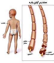
 

سندروم گلين باره


سندروم گلين باره

 (پلي نوريت حاد ايديوپاتيك)                   

تعريف:

سندروم گلين باره يك بيماري نسبتا شايع اعصاب محيطي است كه پس از پوليوميليت شايع ترين علت فلج حاد ژنراليزه است و با دميلينه شدن حاد،سريع و قطعه اي اعصاب در ارتباط است.

اين سندروم با حمله ناگهاني ضعف عضلاني يا فلج مشخص مي شود كه بطور معمول ابتدا در ناحيه پا ايجاد مي شود و در مدت چند روز به سمت بالا گسترش مي يابد و عضلات بازوها،دستان،تنه و صورت را گرفتار مي كند.

اين بيماري هر دو جنس را گرفتار مي كند و متعلق به گروه سني خاصي نيست.

پاتوفيزيولوژي:

اين سندروم در نتيجه واكنش اتوايمون نسبت به ماده ميلين موجود در اعصاب محيطي،نخاعي و جمجمه اي بوجود مي آيد.

در اين سندروم سلول شوآن كه در سيستم عصبي محيطي ميلين توليد مي كند تحليل مي رود و بنابراين امكان دوباره سازي ميلين در فاز بهبودي وجود دارد.

علائم باليني:

آغاز بيماري بصورت حاد يا نيمه حاد بوده و نشانه ها در طول چند روز تا چند هفته كامل مي شود.در ابتدا بيماران بطور شايع از گزگز،مورمور،سوزش و خواب رفتگي اندام ها شكايت دارند(ابتدا پاها و سپس دست ها).بدنبال اختلال حسي،بتدريج ضعف عضلاني متقارن يا نا متقارن به آن افزوده مي شود.

شدت و ضعف بيماري از خفيف تا فلج كامل متغير است.

ساير علائم بصورت دشواري در بلع به علت ضعف عضلات حلق،اختلال در تهويه ريوي به علت فلج عضلات بين دنده اي(وخيم ترين علامت)،فلج شل به علت درگيري نورون هاي حركتي،اختلال حس بصورت پارستزي كه در بخش انتهايي اندام ها بارزتر است،درد بصورت سردرد،كمردرد،تندرنس عضلات اسكلتي در فشار عميق،اختلال دستگاه عصبي اتونوميك بصورت نوسانات شديد در فشارخون سيستوليك،تعريق ناگهاني و شديد،اختلال ادراري،انقباض عروق محيطي،تاكي كاردي حين استراحت و اختلالات هدايت قلب در ECGاست.

همچنين درگيري اعصاب مغزي بطور شايع بصورت فلج زوج هفت يا صورتي و دوبيني تظاهر مي كند.

تشخيص:

علائم و نشانه هاي باليني

افزايش پروتئين و گاماگلوبين در Csf

كاهش قابل توجه در شدت هدايت عصبي در بررسي هاي الكتروفيزيولوژي

قرينه بودن علائم

ميوكايميا(myokymia):حركات ظريف موجي شكل ماهيچه بويژه در ناحيه صورت كه يك نشانه نادر اين سندروم مي باشد.

تاكي كاردي(شايع ترين يافته در ECG)

تغييرات موج T،دپرسيون قطعهST و پهن شدن QRS

درمان:

بيماري پس از شروع در طي يكي دو هفته استقرار مي يابد.

در ماه اول ممكن است نوساناتي در حال عمومي بيمار وجود داشته باشد ولي بعد از 4هفته معمولا سير ثابتي پيدا مي كند و روند بيمار رو به بهبودي مي رود.

درمان عمدتا علامتي است.

بستري شدن در  ICUدر صورت گرفتاري ديافراگم و فلج تنفسي

تهويه مكانيكي ريه ها

لوله گذاري تراشه براي جلوگيري از آسپيراسيون به علت ضعف عضلات حلق

تعويض پلاسما(پلاسما فرز) تجويز ايمونوگلوبولين وريدي

هپارين براي جلوگيري از فلبيت

توان بخشي

نكات بيهوشي:

1.ممكن است وانش هاي جبراني قلبي عروقي وجود نداشته باشد.(افت شديد فشارخون هنگام تغيير وضعيت،خونريزي و..)

2.افزايش شديد فشارخون در اثر تحريكات مثل لارنگوسكوپي به علت بي ثباتي دستگاه اتونوميك

3.خطر آزاد شدن بيش از حد پتاسيم از عضلات فاقد عصب دهي شده بدنبال تجويز اسكولين(پاولن داروي ارجح به علت حداقل آثار سيستميك)

4.نياز به حمايت تنفسي بعد عمل به علت تضعيف ننفس


عوارض سندروم گلين باره:

نوسانات فشارخون

بي نظمي قلبي (تاكي كاردي )

گلومرولونفريت

كاهش غلظت سديم سرم به دليل اختلال ترشح ADH

ادم پاپي به علت افزايش پروتئين مايع نخاع يا احتباس CO2 ناشي از كاهش تهويه ريه

زخم بستر

عواض رواني

دردهاي شديد عضلاني

ترومبوز سياهرگي

آمبولي ريه

پيامد خطرناك :فلج تنفسي و مرگ به دنبال آن

نارسایی قلبی و دیگوکسین

نارسایی احتقانی قلب چیست ؟

شما تنها فردی نمی باشید که مبتلا به نارسایی احتقانی قلب می باشد. افراد بسیاری با نارسایی احتقانی قلب زندگی می کنند. نارسایی احتقانی قلب یکی از شایعترین علل مراجعه به بیمارستان هادر افراد 65 سال یابیشتر می باشد . سالها طول می کشد تا نارسایی قلب ایجاد شود . در نتیجه اگر شماهم اکنون مبتلا به آن نبوده اما در معرض خطر ابتلا به آن هستید ، باید از همین امروز تغییراتی رادر شیوه زندگی خودتان جهت پیشگیری از آن انجام بدهید . 

علایم نارسایی قلبی در طول هفته ها و ماه ها متعاقب ضعیف تر شدن قلب و توانایی کمتر آن برای پمپ کردن خون براساس نیازهای بدن ایجاد می شوند . نارسایی قلب اغلب منجر به بزرگ شدن قلب (بطن چپ) می شود .

آیا قلب شما ایستاده است ؟

ابتلا به نارسایی قلبی به معنی ایستادن قلب از ضربان نمی باشد، بلکه به این مفهوم است که قلب شما به شکل موثری، خون را پمپ نمی کند . قلب کار می کند اما نیاز بدن به خون و اکسیژن تامین نمی شود . نارسایی قلبی در صورت عدم درمان بدتر می شود .رعایت دستورات پزشک بسیار مهم است. اگر شما در شیوه زندگی خودتان تغییرات سالمی را انجام دهید ، احساس بهتری داشته و از زندگی لذت بیشتر ی می برید .

 چه چیز ی ممکن است رخ دهد ؟

 قلب به اندازه کافی خون پمپ نکند .

 خون به سیاهرگها بر گردد .

 مایعات بدن افزایش یابد و منجر به تورم پاها ، مچ پا و ساق پا گردد، که «ادم» نامیده می شود .

 مایعات در بدن تجمع پیدا کنند .

 مایعات در ریه افزایش یابند که به آن «احتقان ریوی» گفته می شود .

 بدن خون ، غذا و اکسیژن به اندازه کافی دریافت نکند .

علایم نارسایی احتقانی قلب چه هستند ؟

 تنگی نفس به ویژه به هنگام دراز کشیدن.

 تورم پاها ، مچ پا و ساق پا.

 اختلالات شناختی یا عدم توانایی تفکر صحیح.

 خستگی ، احساس بی حالی و نزاری.

 افزایش وزن بدلیل احتباس مایع.


علل نارسایی احتقانی قلب چه هستند ؟

 عدم اجازه به جریان خون کافی به قلب ناشی ازسرخرگهای دچار انسداد.

 آسیب به عضله قلبی ناشی از حمله قلبی پیشین .

 وجود نقص قلبی از زمان تولد.

عفونت قلب و یا دریچه های قلب.

پر فشاری خون.

 بیماری دریچه ای قلب.

 بیماری عضلات قلب . 

نارسایی احتقانی قلب چگونه درمان می شود ؟

 استراحت.

مصرف کمتر نمک و غذاهای حاوی نمک.

 ممکن است در برخی موارد،انجام عمل جراحی ضروری باشد .

 ممکن است جهت تقویت قلب شما ،پزشک شما داروهایی تجویز نماید و یا جهت دفع آب اضافی به شما قرص های «دفع آب یا مدر» را بدهد . 

من چه کاری می توانم بکنم ؟

 پیروی از دستورات پزشک .

 مصرف داروها طبق دستورپزشک.

 توزین روزانه خودتان جهت بررسی افزایش مایع.

 مصرف رژیم غذایی سالم حاوی مقادیراندک نمک و چربی اشباع.

من چگونه می توانم بیشتر یاد بگیرم ؟

با پزشک ، پرستار یا مشاور سلامتی خود مشورت کنید . اگر مبتلا به بیماری قلبی می باشید یا قبلا دچار سکته مغزی شده اید، ممکن است اعضای خانواده شما در معرض خطر باشند . ایجاد تغییرات در شیوه زندگی آنها جهت کاهش میزان خطر مهم است .

شما می توانید از کتابچه ها و بروشورهای علمی جهت انتخاب زندگی سالمتر و کاهش میزان خطر ، کنترل بیماری و مراقبت از افراد مورد علاقه تان استفاده کنید .دانایی توانایی است بنابراین بیشتر بیاموزید و بیشتر زندگی کنید.

نارسايي‌ احتقاني‌ قلب‌

توضيح‌ كلي‌

نارسايي‌ احتقاني‌ قلب‌ عبارت‌ است‌ از عارضه‌اي‌ از بسياري‌ از بيماري‌هاي‌ جدي‌ كه‌ طي‌ آن‌ قلب‌ مقداري‌ از ظرفيت‌ پمپ‌ كردن‌ كامل‌ خود را از دست‌ مي‌دهد. بنابراين‌ خون‌ به‌ داخل‌ ديگر اعضاء، خصوصاً ريه‌ها و كبد، پس‌زده‌ مي‌شود.

 علايم‌ شايع‌

تنگي‌ نفس‌، به‌ خصوص‌ هنگام‌ فعاليت‌ يا موقع‌ صاف‌ درازكشيدن‌ روي‌ تخت‌
تند يا نامنظم‌ شدن‌ ضربان‌ قلب‌
برجسته‌ شدن‌ سياهرگ‌هاي‌ گردن‌
خستگي‌، ضعف‌ يا غش‌
سرفه‌ (معمولاً خلط‌دار)
كاهش‌ فشار خون‌
تورم‌ شكم‌، پا و مچ‌ پا
بزرگ‌ شدن‌ كبد

علل‌

حمله‌ قلبي‌؛ بيماري‌ سرخرگ‌هاي‌ قلب‌
بيماري‌ شديد ريه‌ها، مثل‌ آمفيرم‌
كارديوميوپاتي‌ (بيماري‌ عضله‌ قلب‌)
عفونت‌هايي‌ كه‌ به‌ عنوان‌ عارضه‌ يك‌ بيماري‌ زمينه‌اي‌ قلب‌ رخ‌ مي‌دهند.
بيماري‌ دريچه‌هاي‌ قلب‌
ندرتاً تومور قلب‌
فشار خون‌ بالا
نامنظمي‌هاي‌ ضربان‌ قلب‌
پركاري‌ تيروييد
بيماري‌ مادرزادي‌ قلب‌
كم‌خوني‌ شديد


عوامل‌ افزايش‌ دهنده‌ خطر

مصرف‌ الكل‌. الكل‌ باعث‌ كاهش‌ كار قلب‌ مي‌شود.
مصرف‌ بعضي‌ از داروها، از جمله‌ داروهاي‌ مسدودكننده‌ بتا آدرنرژيك‌
عفونت‌هايي‌ كه‌ با تب‌ بالا همراه‌ هستند.
چاقي‌
رژيم‌ پرچرب‌ و پرنمك‌
سيگار كشيدن‌


پيشگيري‌

اگر دچار بيماري‌اي‌ هستيد كه‌ منجر به‌ نارسايي‌ قلب‌ مي‌شود، به‌ پزشك‌ مراجعه‌ كنيد و برنامه‌ درماني‌ مربوطه‌ را به‌ دقت‌ اجرا كرده‌ و بدان‌ پايبند باشيد. دستورات‌ تغذيه‌اي‌ را نيز رعايت‌ كرده‌، كشيدن‌ سيگار و نوشيدن‌ الكل‌ را متوقف‌ كنيد.


عواقب‌ مورد انتظار

اميد به‌ زندگي‌ كاهش‌ مي‌يابد، اما بسياري‌ از موارد را مي‌توان‌ تا مدتي‌ با دارو و گاهي‌ جراحي‌، به‌ خوبي‌ كنترل‌ كرد. در بعضي‌ از موارد مزمن‌ مي‌شود. توجه‌ داشته‌ باشيد كه‌ هرگونه‌ عفونتي‌ باعث‌ بدتر شدن‌ بيماري‌ مي‌شود.


عوارض‌ احتمالي‌

جمع‌ شدن‌ مايع‌ در ريه‌ها
نامنظمي‌هاي‌ ضربان‌ قلب‌


 درمان‌

-امكان‌ دارد آزمايشات‌ تشخيصي‌ شامل‌ موارد زير باشند: آزمايش‌ خون‌ و ادرار؛ اكوكارديوگرافي‌؛ بررسي‌ قلب‌ با كاتتر؛ عكس‌برداري‌ از قلب‌ و ريه‌ها با اشعه‌ ايكس‌؛ آنژيوگرافي‌؛ بررسي‌ راديواكتيو از كارايي‌ عضله‌ قلب ( اسكن‌ تاليم‌)
- هدف‌ از درمان‌ عبارت‌ است‌ بهبود كار پمپ‌ كردن‌ قلب‌، كه‌ معمولاً به‌ كمك‌ دارو، استراحت‌، و ساير اقدامات‌ حمايتي‌ انجام‌ مي‌گيرد.
- گاهي‌ بر حسب‌ علت‌ بيماري‌، ممكن‌ است‌ جراحي‌ روي‌ دريچه‌هاي‌ قلب‌، سرخرگ‌هاي‌ قلب‌، يا آنوريسم‌(شكم‌ آوردن‌) ديواره‌ بطن‌ قلب‌ انجام‌ شود.
- پيوند قلب‌ در موارد شديدي‌ كه‌ به‌ درمان‌ دارويي‌ پاسخ‌ نمي‌دهند.
- دستبند يا گردن‌آويزي‌ همراه‌ خود داشته‌ باشيد كه‌ نوع‌ بيماري‌ و داروهايي‌ كه‌ دريافت‌ مي‌كنيد روي‌ آن‌ نوشته‌ شده‌ باشد.
- بستري‌ شدن‌ در موارد شديد

داروها

داروهاي‌ ادرارآور (ديورتيك‌ها) براي‌ كاهش‌ تجمع‌ مايع‌ در بدن‌ و ورم‌
ديژيتال‌ براي‌ تقويت‌ و تنظيم‌ ضربان‌ قلب‌
داروهاي‌ ضد نامنظمي‌ ضربان‌ قلب‌ براي‌ منظم‌ كردن‌ ضربان‌ قلب‌
داروهاي‌ گشادكننده‌ عروق‌ براي‌ كاهش‌ فشارخون‌، حتي‌ اگر فشارخون‌ طبيعي‌ باشد. اين‌ كار براي‌ برداشتن‌ بار از دوش‌ قلبي‌ كه‌ بايد خون‌ را به‌ درون‌ اين‌ رگ‌ها پمپ‌ كند انجام‌ مي‌گيرد.
اگر از داروهاي‌ ادرارآور يا ديژيتال‌ استفاده‌ مي‌كنيد، بايد پتاسيم‌ بيشتري‌ دريافت‌ بكنيد.


فعاليت‌

در مراحل‌ اوليه‌، استراحت‌ در تخت‌ به‌ حالتي‌ كه‌ قسمت‌ بالاي‌ بدن‌ بالاتر از ساير نقاط‌ بدن‌ قرار گيرد اهميتي‌ به‌ حد دارو دارد. از فعاليت‌ بدني‌ غيرضروري‌(مثل‌ بالارفتن‌ از راه‌ پله‌) تا زماني‌ كه‌ بيماري‌ تحت‌ كنترل‌ در نيامده‌ است‌ خودداري‌ كنيد.
براي‌ كاهش‌ علايم‌، ممكن‌ است‌ لازم‌ شود نوع‌ زندگي‌ خود را تغيير دهيد.


رژيم‌ غذايي‌

سعي‌ كنيد وزن‌ خود به‌ حد ايده‌آل‌ برسانيد تا از فشار روي‌ قلب‌ كاسته‌ شود.
رژيم‌ غذايي‌ بايد كم‌ نمك‌، كم‌ چرب‌، و داراي‌ مقادير زياي‌ فيبر غذايي‌ باشد.
به‌ هيچ‌ عنوان‌ الكل‌ ننوشيد.


در اين‌ شرايط‌ به‌ پزشك‌ خود مراجعه‌ نماييد

اگر شما يا يكي‌ از اعضاي‌ خانواده‌ تان علايم‌ نارسايي‌ احتقاني‌ قلب‌ را داريد.
اگر يكي‌ از موارد زير هنگام‌ درمان‌ رخ‌ دهد:
ـ علايم‌ عفونت‌، از جمله‌ تب‌، دردهاي‌ عضلاني‌، سردرد و سرگيجه‌
ـ بدتر شدن‌ علايم‌، به‌ خصوص‌ تند يا نامنظم‌ شدن‌ ضربان‌ قلب‌ يا بروز خس‌ خس‌ سينه‌ در شب‌
ـ سرفه‌ همراه‌ با زياد شدن‌ خلط‌ يا خوني‌ شدن‌ آن‌
ـ افزايش‌ وزن‌ به‌ ميزان‌ حدود 2- 5/1 كيلوگرم‌ در عرض‌ 2-1 روز
- اگر دچار علايم‌ جديد و غيرقابل كنترل شده ايد. داروهاي‌ مورد استفاده‌ در درمان‌ ممكن‌ است‌ عوارض‌ جانبي‌ به‌ همراه‌ داشته‌ باشند.

همانند بسياري از بيماري هاي ديگر عروق كرونر، ميزان شيوع نارسايي قلبي با افزايش سن زياد مي شود . حداقل 5 ميليون نفر در ايالات متحده دچار نارسایی احتقانی قلب هستند. كه هر سال 550000مورد جديد تشخيص داده مي شود. شيوع آن در افراد بالاي 75سال حدود 10درصد است. بار اقتصادي كه بصورت مستقيم و يا غيرمستقيم توسط نارسايي قلبي ايجاد مي شود. بيش از 25 ميليارد دلار است.

پاتوفيزيولوژي: نارسایی احتقانی قلب مي تواند مجموعه اختلالات قلبي عروقي شامل هايپرتانسيون مزمن ،بيماري عروق كرونر و دريچه اي مي باشد. اين بيماري منجر به كاهش انقباض و پرشدگي مي شود. همانطور كه بيماري قلبي پيشرفت مي كند مكانيسم هاي جبراني نيز فعال شده و مسئول ايجاد علائم و نشانه هايي هستند كه رخ مي دهد.

نارسايي قلبي سيستوليك باعث كاهش حجم خون تخليه شده از بطن مي شود. كاهش كشيدگي بطن توسط بارورسپتور هاي آئورت و اجسام كاروتيد حس مي شود. سپس سيستم عصبي تحريك شده و نوراپي نفرين و اپي نفرين توليد مي كند. كه سبب انقباض عروق مي شودو از عوارض آن كاهش خونرساني به كليه كه كاهش برونده قلب عامل آن است كه فشار خون و پس بار را افزايش مي يابد.

نارسايي قلبي دياستوليك نيز در اثر تداوم باركاري افزايش يافته قلب ايجاد مي شود و بوسيله افزايش تعداد و اندازه ميوكارد پاسخ داده مي شود.كاهش حجم خون در بطن ها سبب كاهش برونده قلبي ميشودو افزايش فشار پر شدگي بطن مي تواند سبب پاسخ هاي عصبي هورموني مي شود كه در نارسايي سيستوليك گفته شد.

اتيولوژي : اختلال عملكرد ميوكارد اغلب به علت بيماري عروق كرونر ،كارديوميوپاتي ،هيپرتانسيون يا اختلالات دريچه اي ايجاد مي شود. بيماران مبتلا به ديابت مليتوس نيز مستعد هستند. آترو اسكلروز شريان هاي كرونر علت اصلي و عمده نارسايي قلبي است. ايسكمي به علت هايپوكسي و اسيدوز ناشي تجمع اسيد لاكتيك سبب اختلال عملكرد ميو كارد مي شود. انفاركتوس ميوكارد نيز سبب نكروز موضعي فلب شده و باعث از دست رفتن قابليت انقباض مي شود.

آموزش به بيمار در مورد دارو  آسپرین: از بيماراني كه تحت درمان طولاني مدت هستند بخواهيد در صورت بروز پتشي و يا خونريزي از لثه يا دستگاه گوارش به پزشك مراجعه كنند.

رژيم غذايي : كم سديم و اجتناب از دريافت مقدار زياد مايع است هدف از محدوديت سديم كاهش مقدار حجم خون در گردش ،كه كاهش دهنده نياز فلب به پمپ اين حجم است. افزايش سديم باعث افزايش مايع در بدن و افزايش بار قلب مي شود . همچنين در طولاني مدت باعث فشار خون مي شود.

تشخيص پرستاري

عدم فعاليت و خستگي مربوط به عدم تعادل بين عرضه و تقاضاي اكسيژن بعلت كاهش برونده

ترك سيگار به خاطر مشكلات قلبي و ريوي

افزايش حجم مايع مربوط به مصرف زياد مايع يا سديم و احتباس مايع مربوط به سندرم نارسايي قلب

كاهش استرس به خاطر نوع زندگي

مداخلات پرستاري

كاهش استرس به خاطر نوع زندگي : —حوادث بیرونی (مانند مشکلات شما با رئیستان، آماده شدن یا نگرانی برای عروسی فرزندتان) می تواند ناراحت کننده باشند . اما به خاطر داشته باشید که این نیروی خارجی نیست که مهم می باشد بلکه واکنش داخلی شما به آن اهمیت دارد . شما نمی توانید تمامی حوادث خارجی زندگی خود را کنترل کنیدولی شما می توانید روش کنترل احساسی و روانی آن ها را تغییر دهید. در اینجا برخی از راه های مطلوب کنار آمدن با آنها ذکر شده اند:

—15 تا 20 دقیقه در روز را به آرامی و ساکت بنشینید، عمیقا نفس بکشید و در مورد یک تصویر آرامش بخش فکر کنید .

— سعی کنید چیزهایی راکه نمی توانید تغییر بدهید را بپذیرید . ضروری نیست که شما همه مشکلات زندگی خود را حل کنید. در مورد مشکلات خود صحبت کنید و به جای شرایط بد به موارد خوب فکر کنید .

— به طور منظم در فعاليت هاي جسماني شرکت کنید . آنچه را که از آن لذت می برید انجام دهید . قدم زدن، شنا ، دوچرخه سواری یا دویدن به منظور فعالیت عضلات بزرگ مناسب می باشند. خارج نمودن تنش ها از بدنتان سبب می شود که احساس بهتری داشته باشید .

—مطابق دستور صریح اسلام الکل مصرف نکنید .در صورت پیروی از سایر ادیان الهی مصرف الکل را محدود کنید. بیش از حد غذا نخورید و سیگار نکشید .

چند توصيه: —در مورد چیزهایی که ممکن است شما را ناراحت کنند، فکر نکنید . ازبرخی چیزها اجتناب کنید مثلا وقت کمتری را با افرادیکه شما را ناراحت می کنند، سپری کنید و یا از رانندگی در ساعات اوج ترافیک پرهیز کنید .

—در مورد مشکلات فکر کنید و سعی کنید آنها را حل کنید . شما می توانید با رئیس خود در مورد مشکلات محل کار صحبت کنید، اگر سگ همسایه شما برای شما مزاحمت ایجاد می کند با وی صحبت کنید و زمانی که کارتان زیاد است از کسی کمک بگیرید .

— روش مواجهه با شرایط دشوار را تغییر دهید. مثبت باشید و نه منفی .

— یاد بگیرید که بگویید «نه». زیاد فکر نکنید. جهت انجام امور به خودتان وقت بیشتری بدهید

—ترك سيگار به خاطر مشكلات قلبي و ريوي

—فواید سلامتی ناشی از ترک سیگار قابل توجه میباشد. تقریباً بلافاصله، گردش خون شخص شروع به بهبود میکند وسطح مونو اکسید کربن در خون شروع به کاهش میکند. مونو اکسید کربن گازی بیرنگ و بی بو است که در دود سیگار یافت میشود و سبب کاهش توانایی خون در حمل اکسیژن می شود. تعداد نبض و فشار خون شخص سیگاری به طور غیر طبیعی بالاست، که پس از ترک سیگار شروع به برگشت به حد طبیعی میکند. بلافاصله چند روز پس از ترک، احساس شخص از مزه و بو برمیگردد و نفس کشیدن برای شخص راحتتر میگردد.

ديگوكسين - دارويي براي قلب

دیگوکسین DIGOXIN 
 
 
دیگوکسین برای افزایش قدرت قلب به عنوان تلمبه تجویز می شود. این دارو با بهبود تعداد ضربان قلب و ضرباهنگ آن چنین تأثیر می گذارد. در نتیجه ، گردش خون بهبود یافته و علایم ناشی از وضعیت بد کارکرد قلب مثل تنگی نفس و تورم دست ها و پاها کاهش می یابد.

چگونگی مصرف
دیگوکسین معمولاً روزی یک بار تجویز می شود. مهم است که دارو هر روز در ساعت یکسانی مصرف شود. معمولاً بهتر است که صبح ها ناشتا خورده شود تا کمتر فراموش شود. البته اگر عادت دارید برای صبحانه غلات برشته بخورید، مراقب باشید دیگوکسین را حداقل یک ساعت قبل یا ۲ ساعت بعد از صبحانه مصرف کنید. (سبوس موجود در این غلات به دیگوکسین متصل شده از جذب آن جلوگیری می کند). هر روز پیش از مصرف دیگوکسین ، نبضتان را به یک دقیقه بشمارید. اگر این کار را بلد نیستید از یک پزشک یا پرستار بخواهید آن را برای شما بیاموزد. اگر نبضتان ۶۰ دقیقه یا کمتر بود، پیش از مصرف داروی آن روز، با پزشکتان مشورت کنید، اگر دیگوکسین مایع مصرف می کنید فقط از قطره چکان مخصوصی که در جعبه دارو موجود است برای اندازه گیری مقدار موردنظر استفاده کنید. از دستورات پزشکتان به دقت پیروی کنید. اگر یک نوبت را فراموش کردید، اگر ظرف ۴ ساعت به یاد آورید مصرفش کنید. در غیراینصورت نوبت فراموش شده را رها کرده به برنامه دارویی معمولتان باز گردید. مقدار دارو را دو برابر نکنید. اگر به هر دلیل دارو را برای دو یا چند نوبت متوالی مصرف نکرده اید پیش از آغاز مجدد دارو با پزشکان مشورت کنید.
هشدارها و عوارض جانبی
درصورت بروز هریک از نشانه های زیر مصرف دیگوکسین را قطع کرده ، و با پزشکتان تماس بگیرید: بی اشتهایی ، اسهال ، تهوع ، استفراغ ، گیجی ، تاری دید (مشاهده یک هاله زرد متمایل به سبز اطراف چراغ ها)، تورم دست ها و پاها، تنگی نفس ، افزایش وزن ناگهانی ۱/۵ کیلوگرم ، در ۲۴ ساعت )، تپش قلب (احساس تکان خوردن شدید قلب در قفسه سینه )، یا نبض زیر ۶۰ در دقیقه .
موارد احتیاط
در صورت وجود هریک از موارد زیر پیش از مصرف دیگوکسین ، پزشکتان را مطلع سازید:
حساسیت به دیگوکسین
باداری یا شیردهی
مصرف داروهای دیگر؛ به ویژه دیگر داروهای دیجیتالیس یا داروهای قلبی دیگر. (آمیودارون ، مسدودکننده های کانال کلسیمی ، پروپافنون ، کینیدین )؛ داروهای پرفشاری خون (مسدودکننده های کانال کلسیمی )؛ مهارکننده های اشتها (قرص های کاهش وزن ، آمفتامین ها)؛ داروهای آسم ، سرفه ، سرماخوردگی ، و سینوزیت ؛ داروهای کاهش کلسترول خون (کلستیرامین ، کلستیپول )؛ داروهای مدر و داروهایی که موجب دفع پتاسیم از بدن می شوند؛ و داروهای حاوی پتاسیم
سابقه یا ابتلا به بیماری های کلیوی
هنگام مصرف دیگوکسین توصیه می شود
به طور مستقیم به پزشکان مراجعه کنید تا بهبودتان را زیر نظر داشته باشد.
یک برگه شناسایی پزشکی همراه داشته باشید که نشان دهد دیگوکسین مصرف می کنید.
دیگوکسین را دور از دسترس کودکان ، و دور از گرما، نور مستقیم ، و حرارت مرطوب نگهداری کنید (در این شرایط دیگوکسین فاسد می شود).
دیگوکسین تاریخ مصرف گذشته را دور از دسترس کودکان در توالت دور بریزید.
قرص های ۲۵% میلی گرم دیگوکسین کوچک و سفید بوده ، شبیه برخی قرص های مدر می باشند. مراقب باشید آنها را با هم اشتباه نکنید. بهتر است هیچگاه قرص های مختلف را داخل یک ظرف مخلوط نکنید.
هنگام مصرف دیگوکسین نباید
شکل دارو، مثلاً قرص و کپسول ، را تغییر دهید. زیرا اگرچه تعداد دارو یکسان باشد، اشکال مختلف دارویی به مقادیر متفاوتی جذب می شوند.
اگر در حال مصرف یک داروی مدر دفع کننده پتاسیم از بدن هستید، مکمل پتاسیم خود را حذف کنید. اگر بدنتان پتاسیم کافی نداشته باشد، ممکن است مسمومیت با دیگوکسین رخ دهد. اگر نمی توانید مکمل پتاسیمی مصرف کنید از پزشکان بخواهید نوع دیگری از دارو را به شما تجویز کند.


کتاب بیهوشی به زبان ساده تألیف دکتر حامد بیضایی

((کتاب بیهوشی به زبان ساده))

تألیف دکتر حامد بیضایی متخصص بیهوشی ومراقبتهای ویژه

منتشر شد

كتاب بيهوشي ميلر مرجعي كامل و معتبر در علم بيهوشي است. كتاب BASIC ميلر نيز به عنوان چكيده‌ای از مطالب آن جهت آشنايي همكاران و فراگيري سريع‌تر بيهوشي كمك بسيار مي‌نمايد. نحوه گفتار و پراكندگي مطالب اين كتاب آن چنان است كه گاه مانع درك صحيح و برداشت درست از آنچه بايد بدانيم، می‌شود.

ممكن است دانشجويان كارشناسي بيهوشي نيز كه از اين كتاب به عنوان مرجع استفاده می‌كنند، با همين مشكل مواجه گردند، لذا ارائه كتابي كه بتواند به راحتي مطالب را در اختيار گذارد، الزامي به نظر مي‌رسيد.

اين كتاب نكات برجسته پنجمين چاپ كتاب BASIC ميلر (چاپ سال 2007 ميلادي) را در بر داشته و حاصل تدريسي است كه در كلاس‌هاي دانشجويان ارائه گرديده است.

براي فهم بهتر اين كتاب، از كتابهاي ديگري همچون Clinical Anesthesiology نوشته G. E. Morgan (چاپ چهارم - 2006 ميلادي)، Nurse Anesthesia Secrets نوشته Mary Karlet (چاپ اول - 2005 ميلادي) و Miller's Anesthesia نوشته Ronald D. Miller (چاپ هفتم – 2010 ميلادي) استفاده گرديده است.

كتاب حاضر براي تمامي افرادي كه در عرصه بيهوشي خدمت می‌نمايند، نوشته شده و در سه جلد ارائه خواهد شد.

جلد اول شامل 13 فصل بوده و مباحث زير را شامل می‌شود:

مقدمه‌ـ تاريخچه‌ـ داروها‌ـ اداره راه هوايي‌ـ بي حسي‌هاي رژيونال (نورآگزيال و محيطي).

در جلد دوم و سوم نيز مباحث زير گنجانده خواهد شد:

فيزيولوژي قلب و ريه‌ـ سيستم اتونوم‌ـ ارزيابي قبل از عمل‌ـ انتخاب نوع بيهوشي‌ـ سيستم‌هاي بيهوشي‌ـ پوزيشن‌ها‌ـ مونيتورينگ‌ـ مايع درماني و ترانسفوزيون‌ـ بيماريهاي قلبي عروقي‌ـ ريوي‌ـ كليوي‌ـ كبدي‌ـ غدد‌ـ CNS‌ـ چشم، گوش، حلق و بيني‌ـ مامايي‌ـ اطفال‌ـ سالمندان‌ـ پيوند عضو‌ـ جراحي سرپايي‌ـ بيهوشي در خارج اتاق عمل‌ـ ريكاوري‌ـ اداره درد‌ـ ICU‌ـ تروما‌ـ احياء قلبي ريوي.

ضمن تشکر از تمامی افرادی که در تهیه این مجموعه یاری نموده‌اند، اميد است مطالعه اين كتاب مورد استفاده همكاران قرار گيرد.

مراقبتهای بیمار پس از اعمال جراحی قلب باز و CABG

مراقبتهای بیمار پس از اعمال جراحی قلب باز و CABG

پس از عمل جراحی بیماران، مستقیما" بیماران به بخش مراقبت های ویژه منتقل می شود. در آنجا بیمار از بی هوشی در می آید. و در آنجا به مدت 24 تا 48 ساعت بستری می شود.در این زمان تعداد زیادی کاتتر و لوله به بیمار متصل است . اهداف پرستاری فوری بعد از عمل جراحی شامل: حفظ تهویه ی کافی مناسب، اکسیژناسیون و ثبات همودینامیکی است. تشخیص و تدابیر سریع برای هر گونه تغییر در وضعیت بیمار امری مهم و حیاتی است. بعلت  اثرات عمل و دستکاری قلب، بیماری تحت جراحی قلب بیش از سایر بیماران جراحی شده دارای وضعیتی بی ثبات و متغیر شده . پرستار باید با نوع عمل انجام شده، دستورات بعد از عمل و بررسی پیشرفته و مشکل گشایی و مهارت ای  تکنیکی که در این بخش ها سر و کار دارد، آشنا باشد.


بیمار بعد از ویزیت بوسیله ی متخصص بی هوشی و بررسی تنفس بیمار بر روی مد SIMV  یا ACV وصل می شود. در بعضی از بیماران، بیمار را بر روی 5 سانتی متر آب PEEP نیز قرار می دهند. پرستار باید از جا گذاری لوله ی هوا اطمینان پیدا کند. و  صدا های تنفسی بیمار را سمع کند.
بعد از عمل، بیمار بای هر چه سریع تر به دشتگاه مانیتورینگ متصل شود تا ریتم قلبی و تعداد ضربان قلب را شناسایی کند. اگر بیمار پیس میکر دارد، نحوه ی کار پیس را ارزیابی کند. سطح پتاسیم خون باید 30 دقیقه بعد از عمل بررسی شود. یک نوار EKG از بیمار گرفته شود.
برای کنترل ثبات همودینامیکی باید CVP بیمار چک شود و اگر نیاز بود از دارو های وازو اکتیو یا اینوتروپیک  استفاده شود.
در مورد  درناژ قفسه ی سینه باید این نکته را توجه داشته باشیم که لوله ی درناژ قفسه ی سینه نباید بیش از اندازه خمیده باشد. باید از بدو ورود و هر یک ساعت یک بار مقدار و کیفیت درناژ را مورد ارزیابی قرار دهیم. هموگلوبین و هماتو  کریت بیمار بیمار باید 30 دقیقه بعد از ورود بیمار به بخش مراقبت های ویژه اندازه گیری شود.
اندازه گیری برون ده ادراری باید هر نیم تا یک ساعت یک بار مورد ارزیابی قرار گیرد. بدلیل رقیق شدن خون بیمار در طی عمل، بیمار حجم ادرارش از 100 تا 200 میلی لیتر افزایش می یابد.  وزن مخصوص ادرار در بیمار کاهش می یابد.باید وجود هر گونه  تیرگی در کیسه ی ادرار را مورد توجه قرار دهیم، زیرا نشانه ی وجود هموگلوبین در ادرار است.


بیمار هنگامی که  به بخش می آید دمای بدن بیمار در حدود 35 تا 36 است، پس پس از عمل باید بیمار را گرم نگهداشت و اندازه گیری شود. از افزایش درجه حرارت بدن به دلیل افزایش متابولیسم و بدن و قلب باید پیشگیری کرد.
بررسی بیمار از لحاظ وضعیت بی هوشی حائز اهمیت است. زیرا بیمار در بدو ورودش به بخش به دلیل اثرات دارو های بی هوشی  فاقد هوشیار است و به تدریج بیدار می شود. در نتیجه باید بیمار را مرتبا" به محیط آگاه کنید  و به قوت قلب دهید.

نوشته شده توسط کاوه جعفری

ارزیابی سطح هوشیاری(GCS)

ارزیابی سطح هوشیاری(GCS)


مقیاس کمایی گلاسکو، ابزاری است برای بررسی پاسخ های بیمار نسبت به محرک ها.
نمرات دامنه ای از اعداد 3(کمای عمیق) تا 15(طبیعی) را در بر می گیرند. در این مقیاس 3 معیار اصلی وجود دارد، که عبارتند از: واکش باز کردن چشم ها، بهترین پاسخ کلامی، و بهترین پاسخ حرکتی به فرامین کلامی و محرک های دردناک.این مقیاس به ویژه برای پایش تغییرات ایجاد شده بعد از آسیب دیدگی های سر و غیره مفید واقع شود، اما نمی تواند جایگزین بررسی جامع و کامل وضعیت عصبی شود.
درواکنش باز کردن به چشم ها می تواند خود بخودی باشد، که نمره ی 4 می گیرد، اگر نسبت به صدا واکنش نشان داد نمره 3، اگر به درد واکنش نشان داد نمره 2، و در صورت عدم واکنش نمره1.
اگر از روی آگاهی، بهترین پاسخ کلامی را داد نمره ی 5، در حالت گیجی نمره ی 4، در صورت استفاده  از لغات نا مناسب، نمره ی 3، صوت های غیر قابل درک نمره ی 2، و به عدم پاسخ نمره ی 1 را می دهیم.
بهترین پاسخ حرکتی: اگر بیمار از دستور اطاعت کرد نمره ی 6، اگر با یک تحریک (درد زا) محل درد را تعیین کرد نمره ی 5، اگر خودش را عقب کشید نمره ی 4، اگر خودش را خم کرد نمره ی 3، و اگر باز کرد نمره ی 2، و اگر نسبت به محرک های درد ناک پاسخی نشان نداد نمره ی 1 را می گیرد.
اگر مجموع نمرات، زیر 7 باشد فرد به کما رفته است. و هر چقدر نمره کمتر باشد، کما عمیق تر است.

در فرم تعیین امتیاز اندازه گیری مردمک ها، تعیین ضربان قلب، تعیین فشار خون، تقارن چشم ها باهم، درجه حرارت و ... هست که باید به آنها هم توجه داشت.


باید توجه داده شود که هر چه میزان GCS پایین تر بیاید، نیاز به عمل جراحی افزایش می یابد.

این مقیاس برای کودکان زیر ۳۶ ماه استفاده نمی شود و از مقیاس PGCS استفاده می شود. در این مقیاس در قسمت کلامی دارای تفاوت تشخیصی برای نمره دهی است. و این تفاوت بخاطر عدم توانایی کودک زیر ۳۶ ماه و عدم تسلط وی در صحبت کردن است و بیشتر تمرکز در این ناحیه بر روی خندیدن و گریه کردن نوزاد و نیز توجه به تحریکات محیطی است.


منبع: پرستاری داخلی جراحی برونر- سودارث 2010ترجمه ی زهرا مشتاق انتشارات جامعه نگر 1389

 نوشته شده توسط کاوه جعفری 


SEVERE ACUTE RESPIRATORY SYNDROME AND INFLUENZA

SEVERE ACUTE RESPIRATORY SYNDROME AND INFLUENZA

“There is no question that there will be another influenza pandemic someday. We simply don't know when it will occur or whether it will be caused by the H5N1 avian influenza virus. It would be prudent to develop robust plans for dealing with such a pandemic.”[72]

Influenza A– and severe acute respiratory syndrome (SARS)–associated viruses are examples of respiratory viruses that may have rampant courses, high virulence, and high lethality. SARS struck like a bolt from the blue in 2002 to 2003 and was a grim reminder of our vulnerability to new infectious diseases. SARS affected mainly people in Asia, in the Pacific Rim, and in Canada. The causative agent is thought to be an RNA coronavirus that is passed on through contact and droplet spread. The virus is viable ex vivo for 24 to 48 hours. Many of the victims of the SARS outbreak were health workers, including anesthesiologists. The influenza pandemic of 1918 to 1919 was one of the major plagues to have affected humankind; it is estimated that Spanish flu left a trail of as many as 25 million corpses around the world in just 25 weeks. A new strain of avian influenza, the H5N1 strain, which is a subtype of influenza A, is now threatening humankind. Influenza is an RNA orthomyxovirus, which, like other RNA viruses, mutates at an alarming rate. The strain H5N1 is so named based on the capsular peptides, hemagglutinin, and neuraminidase. There are 16 known hemagglutinin subtypes and nine known neuraminidase subtypes of influenza A viruses. The Spanish flu was caused by an H1N1 strain of the virus, which continues to cause seasonal human influenza. According to the World Health Organization, pathogenic strains of H5N1 that infect humans may be fatal in approximately 66% of cases. Currently, H5N1 influenza A is passed from bird to human. The fear is that if there is recombination within a patient with concurrent H1N1 or H3N2 influenza A infection, the viruses may undergo recombination resulting in a lethal strain that can spread among humans ( Fig. 19-5 ). Patients with acute respiratory viral infections may require care by anesthesiologists for such procedures as emergency intubation, tracheostomy, chest tube placement, mechanical ventilator support, or general ICU care.


Management of Anesthesia

Preoperative

The anesthesiologist should assess the patient with an appreciation of the potential lethality of the infection. Both patient and their family should be counseled about the high risks associated with SARS-CoV. These viruses are highly contagious and frequently lethal; strict isolation should be enforced and precautions to protect health workers must be taken. The same may apply to a potential (recombinant or newly evolved) influenza strain. Ideally, infected patients should be cared for in rooms with negative pressure to decrease aerosolized spread and contagion. Barrier precautions include the use of full-body disposable oversuits, double gloves, goggles, and powered air-purifying respirators with high-efficiency particulate air filters. [78] [79] If these are not available, N95 masks (block 95% of particles) should be used rather than regular surgical masks. Filters should be placed in both limbs of breathing circuits to protect ventilators and anesthesia machines from contamination. All surfaces should be sterilized with alcohol and rooms should ideally not be used for other patients (if practical) for up to 48 hours after a person with SARS-CoV or H5N1 influenza A has been in the room. Even if it is not SARS or influenza, the same principles of infection control barrier precautions apply to any deadly contagious infection, whether occurring “naturally” or introduced by those with malevolent intent.

Intraoperative

Fears about contagion should not blind anesthesiologists to the high level of care required for these vulnerable patients. During the SARS outbreak in Hong Kong, fears about contagion may have affected patient management.[80] Experience from Canada has shown that when appropriate precautions are taken, spread of infection may be prevented. If mechanical ventilation is required, protective ventilation as for acute respiratory distress syndrome is indicated. Tidal volumes should be limited to 6 to 8 mL/kg lean body mass and mean airway pressure should be less than 30 cm H2O. Sudden cardiorespiratory compromise could be reflective of an expanding pneumothorax. Draining of pleural effusions may improve ventilation and gas exchange. Care should be taken with bronchoscopy as this is a particularly high-risk procedure that results in aerosolization of viral particles.

Signs and Symptoms

Symptoms include nonspecific complaints of viral infection such as cough, sore throat, headache, diarrhea, arthralgia, and muscle pain. In more severe cases, patients may present with respiratory distress, confusion (encephalitis), and hemoptysis. Signs may include fever, tachycardia, sweating, conjunctivitis, rash, tachypnea, use of accessory respiratory muscles, cyanosis, and pulmonary features of pneumonia, pleural effusions, or pneumothorax. A chest radiograph may show patchy infiltrates, areas of opacification, pneumothoraces, and evidence of pleural effusions. Both H5N1 influenza A virus and SARS-coronavirus (CoV) may cause acute lung injury and acute respiratory distress syndrome. Complications include multiorgan failure and severe sepsis


RENAL TRANSPLANTATION

RENAL TRANSPLANTATION

Candidates for renal transplantation are selected from patients with ESRD who are on established programs of long-term hemodialysis. In adults, the most common causes of end-stage renal failure are diabetes mellitus, glomerulonephritis, polycystic kidney disease, and systemic hypertension. Despite concerns about the recurrence of disease in the donor kidney, it has generally been only slowly progressive. A kidney from a cadaver donor can be preserved by perfusion at low temperatures for up to 48 hours, making its transplantation a semielective surgical procedure. Attempts are made to match HLA and ABO blood groups between donor and recipient. Paradoxically, the presence of certain common shared HLA in blood administered to a potential transplant recipient has been observed to induce tolerance to donor antigens and thus improve graft survival. The donor kidney is placed in the lower abdomen and receives its vascular supply from the iliac vessels. The ureter is anastomosed directly to the bladder. Immunosuppressive therapy is instituted during the perioperative period.

Management of Anesthesia

General Anesthesia

Although both regional and general anesthesia have been successfully used for renal transplantation, general anesthesia is most often selected. General anesthesia provides the advantage of mechanically maintaining the patient's ventilation, which may become compromised by surgical retraction in the area of the diaphragm. Drug selection is influenced by known side effects of anesthetic drugs (bowel distention from nitrous oxide, metabolism of sevoflurane to inorganic fluoride). Renal function after kidney transplantation is not predictably influenced by the volatile anesthetic administered. A common approach is to combine volatile anesthetics (isoflurane or desflurane) with nitrous oxide or short-acting opioids. Decreased cardiac output due to negative inotropic effects of volatile anesthetics is minimized to avoid jeopardizing the adequacy of tissue oxygen delivery (especially if anemia is present) and to promote renal perfusion. A high normal systemic blood pressure is required in the presence of euvolemia to maintain adequate urine flow. The selection of muscle relaxants is influenced by the dependence of many of these drugs on renal clearance. In this regard, atracurium, cisatracurium, and mivacurium are attractive selections, as their clearance from the plasma is independent of renal function. A newly transplanted but functioning kidney is able to clear neuromuscular-blocking drugs and the anticholinesterase drugs used for their reversal at the same rate as normal patients.

Central venous pressure monitoring is useful for guiding the rate and volume of crystalloid infusions. Optimal hydration during the intraoperative period is intended to optimize renal blood flow and improve early function of the transplanted kidney. Diuretics are often administered to facilitate urine formation by the newly transplanted kidney. In this regard, osmotic diuretics such as mannitol facilitate urine output and decrease excess tissue and intravascular fluid. Unlike the loop diuretic furosemide, mannitol does not depend on renal tubular concentrating mechanisms to produce diuresis.

When the vascular clamps are released, renal preservative solution from the transplanted kidney and venous drainage from the legs are also released into the circulation. These effluents contain potassium and acid metabolites but, in adults, seem to have minimal systemic effects. Nevertheless, cardiac arrest has been described after completion of the arterial anastomosis to the transplanted kidney and release of the vascular clamp. This event is most likely due to sudden hyperkalemia caused by washout of the potassium-containing preservative solutions from the newly perfused kidney. Unclamping may also be followed by hypotension due to the abrupt addition of up to 300 mL to the capacity of the intravascular fluid space and the release of vasodilating chemicals from previously ischemic tissues. When hypotension results from this change, the treatment is most often intravenous infusion of fluids.

Regional Anesthesia

The advantages of regional anesthesia compared with general anesthesia are the absence of a need for tracheal intubation or administration of neuromuscular blocking drugs. These advantages are negated, however, if regional anesthesia must be extensively supplemented with injected or inhaled drugs. Furthermore, blockade of the peripheral sympathetic nervous system, as produced by regional anesthesia, can complicate control of systemic blood pressure, especially considering the unpredictable intravascular fluid volume status of many of these patients. The use of regional anesthesia, particularly epidural anesthesia, is controversial in the presence of abnormal coagulation.

Postoperative Complications

The newly transplanted kidney may suffer acute immunologic rejection, which manifests in the vasculature of the transplanted kidney. It can be so rapid that inadequate circulation is evident almost immediately after the blood supply to the kidney is established. The only treatment for this acute rejection reaction is removal of the transplanted kidney, especially if the rejection process is accompanied by disseminated intravascular coagulation. A hematoma also may arise in the graft postoperatively, causing vascular or ureteral obstruction.

Delayed signs of graft rejection include fever, local tenderness, and deterioration of urine output. Treatment with high doses of corticosteroids and antilymphocyte globulin may be helpful. The acute tubular necrosis that occurs in the transplanted kidney secondary to prolonged ischemia usually responds to hemodialysis. Cyclosporine toxicity may also cause ARF. Ultrasonography and needle biopsy are performed to differentiate between the possible causes of kidney malfunction.

Opportunistic infections owing to long-term immunosuppression are common after renal transplantation. Long-term survival is unsatisfactory in renal transplant recipients who are immunosuppressed and who also carry hepatitis B surface antigen. The frequency of cancer is 30 to 100 times higher in transplant recipients than in the general population, presumably reflecting the loss of protective effects due to immunosuppression. Large-cell lymphoma is a well-recognized complication of transplantation, occurring almost exclusively in patients with evidence of Epstein-Barr virus infections.


Copyright © 2008 Elsevier Inc. All rights reserved. - www.mdconsult.com

PNEUMONIA

PNEUMONIA

Combined with influenza, community-acquired pneumonia is one of the 10 leading causes of death in the United States. S. pneumoniae is by far the most frequent cause of bacterial pneumonia in adults. Other bacteria that cause pneumonia include H. influenzae, Mycoplasma pneumoniae, S. aureus, Legionella pneumophilia, K. pneumoniae, and Chlamydia pneumoniae. S. pneumoniae usually causes typical pneumonia. Influenzavirus, M. pneumoniae, chlamydia, legionella, adenovirus, and other microorganisms may cause atypical pneumonia.[62]

Diagnosis

An initial chill, followed by abrupt onset of fever, chest pain, dyspnea, fatigue, rigors, cough, and copious sputum production often characterize bacterial pneumonia, although symptoms vary. Nonproductive cough is a feature of atypical pneumonias. A detailed history may suggest possible causative organisms. Hotels and whirlpools are associated with Legionnaires' disease (L. pneumoniae) outbreaks. Fungal pneumonia may occur with cave exploration (Histoplasma capsulatum) and diving (Scedosporium angiospermum). Chlamydia psittaci pneumonia may follow contact with birds and Q fever (Coxiella burnetti) contact with sheep. Alcoholism may increase the risk of bacterial aspiration such as K. pneumoniae. Patients who are immunocompromised, such as those with AIDS, are at risk of fungal pneumonia, such as Pneumocystis jiroveci pneumonia (PCP).

Posteroanterior and lateral chest radiographs may be extremely helpful in diagnosing pneumonia.[63] Diffuse infiltrates are suggestive of an atypical pneumonia whereas a lobar radiographic opacification is suggestive of a typical pneumonia. Atypical pneumonia occurs more frequently in young adults. Radiography is useful for detecting pleural effusions and multilobar involvement. Polymorphonuclear leukocytosis is typical, and arterial hypoxemia may occur in severe cases of bacterial pneumonia. Arterial hypoxemia reflects intrapulmonary shunting of blood owing to perfusion of alveoli filled with inflammatory exudates.

Microscopic examination of sputum plus cultures and sensitivity testing may be helpful in suggesting the etiologic diagnosis of pneumonia and in guiding the selection of appropriate antibiotic treatment. S. pneumoniae and gram-negative organisms, such as H. influenzae, may be seen on sputum stain or culture. Unfortunately, sputum specimens are frequently inadequate and organisms do not invariably grow from sputum. Interpretation of sputum culture may be challenging, as there is frequent normal nasopharyngeal carriage of S. pneumoniae. If there is suspicion, sputum specimens should be sent for acid-fast bacilli (tuberculosis). Antigen detection in urine is a good test for L. pneumophilia. Blood antibody titers are helpful in diagnosing M. pneumoniae. Sputum polymerase chain reaction is useful for chlamydia.[62] Blood cultures are usually negative, but are important to rule our bacteremia. HIV is an important risk factor for pneumonia and should be tested for when pneumonia is suspected.

Management of Anesthesia

Anesthesia and surgery should ideally be deferred with acute infections. Patients with acute pneumonia are often dehydrated and may have renal insufficiency. However, overly aggressive volume resuscitation may worsen gas exchange and morbidity. Fluid management is therefore extremely challenging. Regional anesthesia may be superior. If general anesthesia is unavoidable, a protective ventilation strategy is appropriate with tidal volumes of 6 to 8 mL/kg ideal body mass and mean airway pressures less than 30 cm H2O. The anesthesiologist should suction secretions, send distal sputum specimens for Gram stain and culture, and ensure that appropriate antibiotics are administered both for the pneumonia and to cover the surgery.

Postoperative Pneumonia

Postoperative pneumonia occurs in approximately 20% of patients undergoing major thoracic, esophageal, or major upper abdominal surgery but is rare in other procedures in previously fit patients. Chronic respiratory disease increases the incidence of postoperative pneumonia threefold. Other risk factors include obesity, age older than 70 years, and operations lasting more than 2 hours.[67]

Lung Abscess

Lung abscess may develop after bacterial pneumonia. Alcohol abuse and poor dental hygiene are important risk factors. Septic pulmonary embolization, which is most common in intravenous drug abusers, may also result in formation of a lung abscess. A finding of an air-fluid level on the chest radiograph signifies rupture of the abscess into the bronchial tree, and foul-smelling sputum is characteristic. Antibiotics are the mainstay of treatment of a lung abscess. Surgery is indicated only when complications such as empyema occur. Thoracentesis is necessary to establish the diagnosis of empyema, and treatment requires chest tube drainage and antibiotics. Surgical drainage is necessary to treat chronic empyema.

Copyright © 2008 Elsevier Inc. All rights reserved. - www.mdconsult.com

MANAGEMENT OF HEART FAILURE

MANAGEMENT OF HEART FAILURE

Current therapeutic strategies are aimed at reversing the pathophysiologic alterations present in heart failure and at interrupting the vicious circle of maladaptive mechanisms ( Fig. 6-3 ). Short-term therapeutic goals in patients with heart failure include relieving symptoms of circulatory congestion, increasing tissue perfusion, and improving the quality of life. However, management of heart failure involves more than the treatment of symptoms. The processes that contributed to the LV dysfunction may progress independently of the development of symptoms. Therefore, the long-term therapeutic goal is to prolong life by slowing or reversing the progression of ventricular remodeling.

Picture Hosted by Free Photo Hosting at <A href="http://www.iranxm.com/

Figure 6-3  Primary targets of treatment in heart failure. Treatment options for patients with heart failure affect the pathophysiologic mechanisms that are stimulated in heart failure. Angiotensin-converting enzyme inhibitors (ACEIs) and angiotensin II receptor blockers decrease afterload by interfering with the renin-angiotensin-aldosterone system, resulting in peripheral vasodilation. They also affect left ventricular hypertrophy, remodeling, and renal blood flow. Aldosterone production by the adrenal glands is increased in heart failure. It stimulates renal sodium retention and potassium excretion and promotes ventricular and vascular hypertrophy. Aldosterone antagonists counteract the many effects of aldosterone. Diuretics decrease preload by stimulating natriuresis in the kidneys. Digoxin affects the Na+K+ATPase pump in the myocardial cell, increasing contractility. Inotropes such as dobutamine and milrinone increase myocardial contractility. β-Blockers inhibit the sympathetic nervous system and adrenergic receptors. They slow the heart rate, decrease blood pressure, and have a direct beneficial effect on the myocardium by enhancing reverse remodeling. Selected agents that also block α-adrenergic receptors can cause vasodilation. Vasodilator therapy such as combination therapy with hydralazine and isosorbide dinitrate decreases afterload by counteracting peripheral vasoconstriction. Cardiac resynchronization therapy with biventricular pacing improves left ventricular function and favors reverse remodeling. Nesiritide (brain natriuretic peptide) decreases preload by stimulating diuresis and decreases afterload by vasodilation. Exercise improves peripheral blood flow by eventually counteracting peripheral vasoconstriction. It also improves skeletal-muscle physiology.  (Reproduced with permission from Jessup M, Brozena S: Heart failure. N Engl J Med 2003;348:2007–2018. Copyright © 2003 Massachusetts Medical Society. All rights reserved.)


Management of Chronic Heart Failure

The current recommended therapy of chronic heart failure is based on results of large, adequately powered, randomized trials and on the American College of Cardiology/American Heart Association and European Society of Cardiology guidelines for the diagnosis and treatment of chronic heart failure. According to these guidelines, treatment options include lifestyle modification, patient and family education, medical therapy, corrective surgery, implantable devices, and cardiac transplantation ( Fig. 6-4 ).

Picture Hosted by Free Photo Hosting at <A href="http://www.iranxm.com/

Figure 6-4  Stages of heart failure and treatment options for systolic heart failure. Patients with stage A heart failure are at high risk of heart failure but do not yet have structural heart disease or symptoms of heart failure. This group includes patients with hypertension, diabetes, coronary artery disease, previous exposure to cardiotoxic drugs, or a family history of cardiomyopathy. Patients with stage B heart failure have structural heart disease but no symptoms of heart failure. This group includes patients with left ventricular hypertrophy, previous myocardial infarction, left ventricular systolic dysfunction, or valvular heart disease, all of whom would be considered to have New York Heart Association (NYHA) class I symptoms. Patients with stage C heart failure have known structural heart disease and current or previous symptoms of heart failure. Their current symptoms may be classified as NYHA class I, II, III, or IV. Patients with stage D heart failure have refractory symptoms of heart failure at rest despite maximal medical therapy, are hospitalized, and require specialized interventions or hospice care. All such patients would be considered to have NYHA class IV symptoms. ACE, angiotensin-converting enzyme; ARB, angiotensin receptor blocker; VAD, ventricular assist device.  (Reproduced with permission from Jessup M, Brozena S: Heart failure. N Engl J Med 2003;348:2007–2018. Copyright © 2003 Massachusetts Medical Society. All rights reserved.)


Lifestyle modifications are aimed at decreasing the risk of heart disease and include smoking cessation, a healthy diet with moderate sodium restriction, weight control, exercise, moderate alcohol consumption, and adequate glycemic control.

Copyright © 2008 Elsevier Inc. All rights reserved. - www.mdconsult.com


Management of Acute Heart Failure

Management of Acute Heart Failure

Patients may experience acute heart failure as a result of decompensated chronic heart failure or de novo. Anesthesiologists deal with acute heart failure when caring for patients in overt heart failure who present for emergency surgery or patients who decompensate intraoperatively. Acute heart failure therapy has three phases: the emergency phase, the in-hospital management phase, and the predischarge phase. For the anesthesiologist, the emergency phase is of most interest and is the phase that is addressed here. The hemodynamic profile of acute heart failure is characterized by high ventricular filling pressures, low cardiac output, and hyper- or hypotension. Traditional therapy includes diuretics, vasodilators, inotropic drugs, mechanical assisted devices (intra-aortic balloon pump, ventricular assist device), and emergency cardiac surgery. Newer therapy includes calcium sensitizers, exogenous BNP, and nitric oxide synthase inhibitors.

Diuretics and Vasodilators

Loop diuretics can improve symptoms rapidly, but in high doses, they may have deleterious effects on clinical outcomes. It may be more desirable to use a combination of a low dose of loop diuretic with an intravenous vasodilator. Nitroglycerin and nitroprusside reduce LV filling pressure and systemic vascular resistance and increase stroke volume. However, nitroprusside may have a negative impact on clinical outcome in patients with acute myocardial infarction.

Inotropic Support

Positive inotropic drugs have been the mainstay of treatment for patients in cardiogenic shock. Their positive inotropic effect is produced via an increase in cyclic adenosine monophosphate, which promotes an increase in intracellular calcium levels and, thereby, an improvement in excitation-contraction coupling. Catecholamines (epinephrine, norepinephrine, dopamine, and dobutamine) do so by direct β-receptor stimulation, whereas phosphodiesterase inhibitors (amrinone, milrinone) block the degradation of cyclic adenosine monophosphate. Side effects of inotropic drugs include tachycardia, increased myocardial energy demand and oxygen consumption, dysrhythmias, worsening of DHF, and down-regulation of β-receptors. Long-term use of these drugs may result in cardiotoxicity and accelerate myocardial cell death.

Calcium Sensitizers

Myofilament calcium sensitizers are a new class of positive inotropic drugs that increase contractility without increasing intracellular levels of calcium. Therefore, there is no significant increase in myocardial oxygen consumption or heart rate and no propensity for dysrhythmias. The most widely used medication in this class is levosemindan. It is an inodilator increasing myocardial contractile strength and promoting dilation of systemic, pulmonary, and coronary arteries. It does not worsen diastolic function. Studies have shown that levosemindan may be particularly useful in the setting of myocardial ischemia. Levosemindan is included in the European guidelines for treatment of acute heart failure, but it is not yet available for use in the United States.

Exogenous B-Type Natriuretic Peptide

Nesiritide (Natrecor) is recombinant BNP that binds to both the A- and B-type natriuretic receptors. It promotes arterial, venous, and coronary vasodilation, thereby decreasing LVEDP and improving dyspnea. Nesiritide induces diuresis and natriuresis. It has many effects similar to nitroglycerin but generally produces less hypotension and more diuresis than nitroglycerin.

Nitric Oxide Synthase Inhibitors

The inflammatory cascade stimulated by heart failure results in production of a large amount of nitric oxide in the heart and vascular endothelium. These high levels of nitric oxide have a negative inotropic and profound vasodilatory effect leading to cardiogenic shock and vascular collapse. Inhibition of nitric oxide synthase should decrease these harmful effects. L-NAME (N-nitro-L-arginine methyl ester) is the principal drug in this class under investigation.

Mechanical Devices

If the etiology of acute heart failure is a large myocardial infarction, the insertion of an intra-aortic balloon pump should be considered. The intra-aortic balloon pump is a mechanical device inserted via the femoral artery and positioned just below the left subclavian artery. Its balloon inflates in diastole increasing aortic diastolic blood pressure and coronary perfusion pressure. The balloon deflates in systole creating a “suction” effect that enhances LV ejection. Complications of intra-aortic balloon pump placement include femoral artery or aortic dissection, bleeding, thrombosis, and infection.

In severe cardiogenic shock, emergency insertion of LV and/or right ventricular assist devices may be necessary for survival.

Copyright © 2008 Elsevier Inc. All rights reserved. - www.mdconsult.com


EPIDERMOLYSIS BULLOSA

EPIDERMOLYSIS BULLOSA

Epidermolysis bullosa is a group of genetic diseases of mucous membranes and skin, particularly the oropharynx and esophagus. Epidermis bullosa can be categorized as simplex, junctional, and dystrophic. In the simplex type, epidermal cells are fragile and mutations of genes encoding keratin intermediate filament proteins underlie the fragility. In the dystrophic types (incidence approximately one in every 300,000 births), the genetic mutation appears to be in the gene encoding the type of collagen that is the major component of anchoring fibrils

Management of Anesthesia

Supplemental corticosteroids may be indicated during the perioperative period if patients have been on long-term treatment with these drugs. The main anesthetic concerns in patients with epidermolysis bullosa center on the serious complications that can occur if proper precautions are not taken during instrumentation. Avoidance of trauma to the skin and mucous membranes is crucial. Bulla formation can be caused by trauma from tape, blood pressure cuffs, tourniquets, adhesive electrodes, and rubbing the skin with alcohol wipes. Blood pressure cuffs should be padded with a loose cotton dressing. Electrodes should have the adhesive portion removed. Petroleum jelly gauze can help hold them in place. Anything that touches a patient should be well padded. Intravenous and intra-arterial catheters should be sutured or held in place with gauze wraps rather than tape. A nonadhesive pulse oximetry sensor should be used. A soft foam, sheepskin, or gel pad should be placed under the patient. All creases should be removed from the linen.

Trauma from the anesthetic face mask must be minimized by gentle application against the face. Lubrication of the face and mask with cortisol ointment, or indeed any lubricant, can be helpful. Upper airway instrumentation should be minimized because the squamous epithelium lining the oropharynx and esophagus is very susceptible to trauma. Frictional trauma to the oropharynx, such as that produced by an oral airway, can result in formation of large intraoral bullae and/or extensive hemorrhage from denuded mucosa. Nasal airways are equally hazardous. Esophageal stethoscopes should be avoided. Hemorrhage from ruptured oral bullae has been treated successfully with epinephrine-soaked gauze applied directly to the bullae.

Interestingly, endotracheal intubation has not been associated with laryngeal or tracheal complications in patients with epidermolysis bullosa dystrophica. Indeed, laryngeal involvement with this form of the disease is rare, and tracheal bullae have not been reported. This finding is consistent with the greater resistance of columnar epithelium to disruption compared to fragile squamous epithelium. Generous lubrication of the laryngoscope blade with cortisol ointment and/or petroleum jelly and selection of a smaller endotracheal tube than usual are recommended. Chronic scarring of the oral cavity can result in a narrow oral aperture and immobility of the tongue making tracheal intubation difficult. After intubation, the tube must be carefully immobilized with soft cloth bandages to prevent movement in the oropharynx, and the tube must be positioned so that it does not exert lateral forces at the corners of the mouth. Tape is not used to hold the endotracheal tube in place. It must be remembered that oropharyngeal suctioning can lead to life-threatening bulla formation. The risk of pulmonary aspiration may be increased in the presence of esophageal stricture.

Porphyria cutanea tarda has been reported to occur with increased frequency in patients with epidermolysis bullosa. This type of porphyria does not have the same implications for management of anesthesia, as does acute intermittent porphyria.

Propofol and ketamine are useful for avoiding airway manipulation when the operative procedure does not require controlled ventilation or skeletal muscle relaxation. Despite the presence of dystrophic skeletal muscle, there is no evidence that these patients are at increased risk of a hyperkalemic response when treated with succinylcholine. There are no known contraindications to the use of volatile anesthetics in these patients. As alternatives to general anesthesia, regional anesthetic techniques (spinal, epidural, brachial plexus block) have been recommended.

Copyright © 2008 Elsevier Inc. All rights reserved. - www.mdconsult.com


Colorectal Cancer

Colorectal Cancer

Colon cancer is second only to lung cancer as a cause of cancer death in the United States. The incidence and mortality from this cancer has not changed appreciably during the past several decades. Almost all colorectal cancers are adenocarcinomas, and the disease generally occurs in adults older than 50 years.

Etiology

Most colorectal cancers arise from premalignant adenomatous polyps. Large polyps, especially those larger than 1.5 cm in diameter, are more likely to contain invasive cancer. Although adenomatous polyps are common (present in more than 30% of patients older than 50 years), less than 1% of adenomatous polyps ever become malignant. It is thought that adenomatous polyps require at least 5 years of growth before they become clinically significant. The evolution of normal colonic mucosa to a benign adenomatous polyp that contains cancer and then to life-threatening invasive cancer is associated with a series of genetic events that involve the mutational activation of a proto-oncogene and the loss of several genes that normally suppress tumorigenesis.

Most colorectal cancers appear to be related to diet, with the disease occurring in the greatest incidence among individuals in upper socioeconomic classes living in urban areas. There is a direct correlation between calories consumed, dietary fat and oil, and meat protein. Available data indicate that a high intake of animal fat is the dietary element that is most strongly associated with the risk of colon cancer. As many as 25% of patients with colorectal cancer have a family history of the disease. Inflammatory bowel disease is associated with an increased incidence of colorectal cancer. Cigarette smoking for longer than 35 years appears to increase the risk of colorectal cancer.

Diagnosis

The rationale for colorectal cancer screening is that early detection and removal of localized superficial tumors and precancerous lesions in asymptomatic individuals increases the cure rate. Screening programs (digital rectal examination, examination of the stool for occult blood, colonoscopy) appear to be particularly useful for persons who have first-degree relatives with a history of the disease, especially if these relatives developed the colorectal cancer before 55 years of age. There is evidence that annual or biennial fecal occult blood testing is associated with a decreased incidence of colorectal cancer.

Signs and Symptoms

The presenting signs and symptoms of colorectal cancer reflect the anatomic location of the cancer. Because stool is relatively liquid as it passes into the right colon through the ileocecal valve, tumors in the cecum and ascending colon can become large and markedly narrow the bowel lumen without causing obstructive symptoms. Ascending colon cancers frequently ulcerate, leading to chronic blood loss in the stool. These patients experience symptoms related to anemia, including fatigue and, in some patients, angina pectoris.

Stool becomes more concentrated as it passes into the transverse colon. Transverse colon cancers cause abdominal cramping, occasional bowel obstruction, and even perforation. Abdominal radiographs reveals characteristic abnormalities in the colonic gas pattern, reflecting narrowing of the lumen (“napkin ring lesion”). Colon cancers developing in the rectosigmoid portion of the large intestine result in tenesmus and thinner stools. Anemia is unusual despite the passage of bright red blood from the rectum (often attributable to hemorrhoids).

Colorectal cancers initially spread to regional lymph nodes and then through the portal venous circulation to the liver, which represents the most common visceral site of metastases. Colorectal cancers rarely spread to lung, bone, or brain in the absence of liver metastases. A preoperative increase in the serum concentration of carcinoembryonic antigen suggests that the tumor will recur following surgical resection. Carcinoembryonic antigen is a glycoprotein that is also increased in the presence of other cancers (stomach, pancreas, breast, lung) and nonmalignant conditions (alcoholic liver disease, inflammatory bowel disease, cigarette smoking, pancreatitis).

Treatment

The prognosis for patients with adenocarcinoma of the colorectum depends on the depth of tumor penetration into the bowel wall and the presence or absence of regional lymph node involvement and distant metastases (liver, lung, bone). Radical surgical resection, which includes the blood vessels and lymph nodes draining the involved bowel, offers the best potential for cure. Surgical management of cancers that arise in the distal rectum may necessitate a permanent sigmoid colostomy (abdominoperineal resection). Because most recurrences occur within 3 to 4 years, the cure rate for colorectal cancer is often estimated by 5-year survival rates.

Radiation therapy is a consideration in patients with rectal tumors since the risk of recurrence following surgery is significant. Postoperative radiation therapy causes transient diarrhea and cystitis, but permanent damage to the small intestine and bladder is uncommon. Use of chemotherapy in patients with advanced colorectal cancers rarely results in a satisfactory response.

Management of Anesthesia

Management of anesthesia for surgical resection of colorectal cancers may be influenced by anemia and the effects of metastatic lesions in liver, lung, bone, or brain. Chronic large bowel obstruction probably does not increase the risk of aspiration during induction of anesthesia, although abdominal distention could interfere with adequate ventilation and oxygenation. Blood transfusion during surgical resection of colorectal cancers has been alleged to be associated with a decrease in the length of patient survival. This could reflect immunosuppression produced by transfused blood. For this reason, careful review of the risks and benefits of blood transfusions in these patients is prudent.

Copyright © 2008 Elsevier Inc. All rights reserved. - www.mdconsult.com

CIRRHOSIS

CIRRHOSIS

Cirrhosis can result from a large variety of chronic, progressive liver diseases. Most often cirrhosis is the result of excessive chronic alcohol ingestion or chronic viral hepatitis due to HBV or HCV infection. Scarring of the liver results in disruption of normal liver architecture, and regenerating parenchymal nodules are typically seen. The pattern of scarring seldom permits determination of a specific etiology, but other histologic features may provide clues as to the cause of the cirrhosis.

Diagnosis

Percutaneous liver biopsy establishes the diagnosis of cirrhosis. Computed tomography, magnetic resonance imaging, and hepatic ultrasonography with Doppler flow studies may reveal findings consistent with cirrhosis (splenomegaly, ascites, irregular liver surface). Upper gastrointestinal endoscopy can establish the presence of esophagogastric varices.

Management of Anesthesia

It is estimated that 5% to 10% of patients with cirrhosis require surgery in the last 2 years of life. Many trauma beds are occupied by patients who were injured while under the influence of alcohol. In those patients who abuse alcohol, the presence of ascites, sepsis, and chronic obstructive pulmonary disease preoperatively is associated with increased postoperative morbidity and mortality. Postoperative morbidity includes pneumonia, bleeding, sepsis, poor wound healing, and deterioration in liver function. The pathogenic mechanisms of these complications often includes subclinical cardiorespiratory insufficiency and immune incompetence. The complications of alcohol withdrawal can also affect perioperative morbidity.

Preoperative Preparation

Certain preoperative criteria correlate with surgical risk and postoperative outcome in patients with cirrhosis undergoing major surgery ( Table 11-6 ). Identifying co-existing problems that can be optimized preoperatively (cardiorespiratory function, coagulation status, renal function, intravascular fluid volume, electrolyte balance, nutrition) may decrease morbidity and mortality associated with elective surgery in patients with severe liver disease. Coagulation status should be evaluated and parenteral vitamin K administered if the prothrombin time is prolonged. Failure of parenteral vitamin K to improve synthesis of prothrombin suggests the presence of severe hepatocellular disease. Impaired prothrombin production due to biliary obstruction and the absence of bile salts that facilitate gastrointestinal absorption of vitamin K is promptly reversed by parenteral vitamin K therapy. Thrombocytopenia, which often accompanies severe liver disease, may require treatment. Hypoglycemia may be present, and administration of a glucose solution is a consideration perioperatively. There should be proper hydration and urine output prior to surgery. Hepatic blood flow is predictably decreased in patients with cirrhosis, and any further decrease due to anesthetic-induced depression of cardiac output or blood pressure could jeopardize hepatocyte oxygenation.


TABLE 11–6   -- Prediction of Perioperative Risk in the Patient with Liver Disease

Parameter

Low Risk

Moderate Risk

High Risk

Bilirubin (mg/dL)

<2

2–3

>3

Albumin (g/dL)

>3.5

3.0–3.5

<3

Prothrombin time (seconds prolonged)

1–4

4–6

>6

Encephalopathy

None

Moderate

Severe

Nutrition

Excellent

Good

Poor

Ascites

None

Moderate

Marked

Adapted from Strunin I: Preoperative assessment of the patient with liver dysfunction. Br J Anaesth 1978;50:25–34.


Chronic alcohol ingestion has been demonstrated to increase anesthetic requirements (MAC) for isoflurane most likely due to cross-tolerance. Accelerated metabolism of drugs in the presence of alcohol-induced microsomal enzyme induction can also alter the amount of anesthetic drug needed to achieve a certain anesthesia depth. Decreased protein binding of drugs in the presence of hypoalbuminemia could increase the pharmacologically active fraction of intravenous anesthetic drugs. Alcohol-induced cardiomyopathy could make patients unusually sensitive to the cardiac depressant effects of volatile anesthetics. There may be decreased responsiveness to catecholamines.

Intoxicated Alcoholic Patients

In contrast to a chronic but sober alcoholic patient, the acutely intoxicated patient requires less anesthetic because there are additive depressant effects from the alcohol and the anesthetic drugs. Acutely intoxicated patients are also ill equipped to withstand stress and acute surgical blood loss. Furthermore, alcohol may decrease the tolerance of the brain to hypoxia. Intoxicated patients may be more vulnerable to regurgitation of gastric contents since alcohol delays gastric emptying and decreases lower esophageal sphincter tone. Surgical bleeding may reflect alcohol-induced interference with platelet aggregation. Alcohol, even in moderate doses, causes increased plasma catecholamine concentrations, most likely reflecting inhibition of neurotransmitter uptake back into presynaptic nerve endings.

Intraoperative Management

Optimal anesthetic drug choices or techniques in the presence of liver disease are unknown. It is important to remember, however, that a constant feature of chronic liver disease is decreased hepatic blood flow due to portal hypertension. As a result, hepatic blood flow and hepatocyte oxygenation are more dependent on hepatic artery blood flow than normally. The hepatic artery may provide more than 50% of the oxygen supply by vasodilating during periods of decreased portal vein blood flow. Hepatic blood flow and hepatocyte oxygenation seem to be well maintained during administration of isoflurane, desflurane, and sevoflurane but not halothane. However, the ability of the hepatic artery to vasodilate in response to a decrease in portal vein blood flow can be blunted by volatile anesthetics, especially halothane and especially in high concentrations. It is prudent to limit the dose of volatile anesthetic to minimize the likelihood of a persistent decrease in mean arterial pressure because intraoperative hypotension may be associated with increased postoperative morbidity and mortality. Intravenous anesthetic drugs are valuable adjuncts to volatile anesthetics with or without nitrous oxide, but cumulative drug effects are likely if liver disease is severe enough to slow metabolism of the intravenous anesthetics. Regardless of the drugs selected for anesthesia, postoperative liver dysfunction is likely to be exaggerated in patients with chronic liver disease, presumably due to the effects of anesthetic drugs and/or stress-induced activation of the sympathetic nervous system on hepatocyte oxygenation. Regional anesthesia can be useful in patients with advanced liver disease if the coagulation status is acceptable.

Muscle Relaxants

Hepatic clearance of muscle relaxants must be considered when selecting a particular neuromuscular blocker for administration to patients with cirrhosis. Succinylcholine or mivacurium are acceptable, although severe liver disease may decrease plasma cholinesterase activity and prolong the duration of action of these drugs. The increased volume of distribution that accompanies cirrhosis, especially with ascites, will result in the need for a larger initial dose of nondepolarizing muscle relaxant to produce the required plasma concentration. However, subsequent doses may be smaller due to decreased hepatic clearance and metabolism. Hepatic dysfunction does not alter the elimination half-time of atracurium or cisatracurium. The elimination half-time of vecuronium is not increased until the dose exceeds 0.1 mg/kg, consistent with the dependence of this drug on hepatic clearance. Altered protein binding of muscle relaxants is insignificant as a mechanism of an altered response in patients with cirrhosis.

Monitoring

Monitoring of arterial blood gases and urine output is often necessary. The need for invasive intraoperative monitoring is determined by the extent and urgency of the surgery. Management of anesthesia for surgical creation of a portocaval shunt includes monitoring arterial pressure and cardiac filling pressures. Fluid administration must be carefully titrated to an endpoint such as central venous pressure, pulmonary artery occlusion pressure, and urine output. Intraoperative maintenance of an acceptable urine output may help decrease the risk of postoperative acute renal failure. When blood replacement is necessary, the stored blood should be administered as slowly as possible to compensate for the decreased clearance of citrate by the cirrhotic liver. Infusion of glucose may be necessary during the perioperative period to prevent hypoglycemia. A practical point is avoidance of unnecessary esophageal instrumentation (esophageal stethoscope, orogastric or nasogastric tube) in patients with known esophageal varices.

Postoperative Management

Regardless of the drugs selected for anesthesia, postoperative liver dysfunction/jaundice is likely in patients with chronic liver disease. Cholestasis and sepsis can also be causes of postoperative jaundice. Manifestations of alcohol withdrawal usually appear 24 to 72 hours after cessation of drinking and can constitute a medical emergency in the postoperative period.

Copyright © 2008 Elsevier Inc. All rights reserved. - www.mdconsult.com

ACUTE LIVER FAILURE

ACUTE LIVER FAILURE

Acute hepatic failure is characterized by jaundice, hypoalbuminemia, coagulopathy, malnutrition, susceptibility to infection, and renal dysfunction in the clinical setting of acute hepatic disease. Fulminant hepatic failure refers to acute liver failure with superimposed hepatic encephalopathy that develops within 2 to 8 weeks of the onset of illness in a patient without preexisting liver disease. Viral hepatitis and drug-induced liver injury account for most cases of acute liver failure

Management of Anesthesia

Only surgery necessary to correct life-threatening problems should be considered in patients with acute liver failure. Preoperative correction of coagulation abnormalities with fresh frozen plasma may be indicated. Low doses of volatile anesthetics or even nitrous oxide alone may be sufficient to provide analgesia and amnesia in these critically ill patients. Intravenous anesthetics may have prolonged effects due to the marked reduction in metabolism of these drugs. Muscle relaxants may be needed to facilitate operative exposure or manage ventilation. When choosing a muscle relaxant, one must consider the impact of decreased hepatic function and associated renal dysfunction on the clearance of the drug. Because the plasma half-life of pseudocholinesterase is 14 days, it is unlikely that acute liver failure would be associated with a prolonged response to succinylcholine or mivacurium.

Administration of glucose is important, and plasma glucose measurement to confirm the absence of hypoglycemia is prudent. Blood should be administered as slowly as possible to minimize the likelihood of citrate intoxication. Monitoring arterial blood gases and electrolyte concentrations is helpful since these patients are vulnerable to development of arterial hypoxemia, metabolic acidosis, hypokalemia, hypocalcemia, and hypomagnesemia. Hypotension and its adverse effect on hepatic blood flow and hepatocyte oxygenation must be considered. Urine output is maintained with intravenous infusion of crystalloid or colloid and, if necessary, diuretic administration. Invasive monitoring is helpful for guiding overall hemodynamic management. These patients are vulnerable to infection, emphasizing the importance of aseptic technique during insertion of intravascular catheters. Lactulose therapy during the preoperative period may decrease the ammonia load and help prevent development of hepatic encephalopathy.

Copyright © 2008 Elsevier Inc. All rights reserved. - www.mdconsult.com


دانلود فایل پاورپوینت هوشبرهای داخل وریدی

http://www.faupload.com/upload/90/Dey/1-15/هوشبرهای-داخل-وريدی.pptx

دانلود فایل پاورپوینت روش های بیهوشی

http://www.faupload.com/upload/90/Dey/1-15/روشهای-بیهوشی.pptx

دانلود فایل پاورپوینت ترانسفوزیون خون

http://www.faupload.com/upload/90/Dey/1-15/TRasfusion.pptx

دانلود فایل پاورپوینت داروهای مخدر

http://www.faupload.com/upload/90/Dey/1-15/Opioids.pptx

دانلود فایل پاورپوینت داروهای شل کننده بیهوشی

http://www.faupload.com/upload/90/Dey/1-15/NMB-Drugs-2-.pptx

دانلود فایل پاورپوینت سیستم های مپلسون

http://www.faupload.com/upload/90/Dey/1-15/Mapleson-system.pptx

دانلود فايل پاورپوينت آشنايي با بخش مراقبتهاي ويژه

http://www.faupload.com/upload/90/Aban/20-26/ICU.pptx

دانلود فايل پاورپوينت اكسيژن تراپي

http://www.faupload.com/upload/90/Aban/20-26/اكسي‍ژن-تراپي.pptx

دانلود فايل پاورپوينت مانيتورينگ هموديناميك

http://www.faupload.com/upload/90/Aban/20-26/Hemodinamic.pptx

دانلود فايل پاورپوينت آناتومی و فیزیولوژی راه هاي هوايي

http://www.faupload.com/upload/90/Aban/20-26/ANATOMY.pptx

دانلود فايل پاورپوينت مراقبتهاي پرستاري از لوله تراشه

http://www.faupload.com/upload/90/Aban/20-26/Loleh-trashe.pptx

دانلود فايل پاورپوينت آناليز گازهاي خون شرياني

http://www.faupload.com/upload/90/Aban/20-26/gasometry.pptx

دانلود فایل پاورپوینت ونتیلاسیون مکانیکی

http://s1.faupload.com/do.php?filename=8fa_88-mechanical-ventilation.ppt

دانلود فایل پاورپوینت مدهای تنفسی در ونتیلاتورها

http://www.faupload.com/upload/90/Aban/20-26/MODE.pptx

دانلود فایل پاورپوینت منحنی های تنفسی در ونتیلاتور

http://s1.faupload.com/do.php?filename=c19_منحنی-های-تنفسی-در-ونتیلاتور.pptx